
Page 326 - Read Online
P. 326
Page 2 of 10 Yao et al. Hepatoma Res 2018;4:30 I http://dx.doi.org/10.20517/2394-5079.2018.32
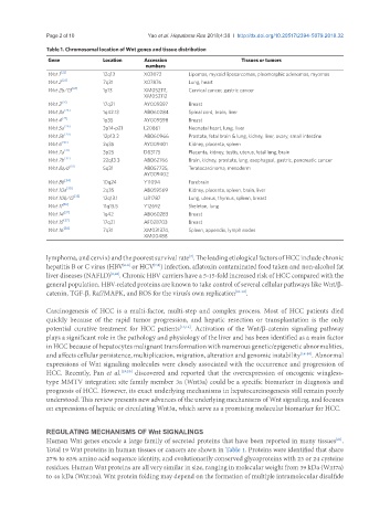
Table 1. Chromosomal location of Wnt genes and tissue distribution
Gene Location Accession Tissues or tumors
numbers
Wnt 1 [22] 12q13 X03072 Lipomas, myxoid liposarcomas, pleomorphic adenomas, myomas
Wnt 2 [23] 7q31 X07876 Lung, heart
Wnt 2b/13 [24] 1p13 XM052111, Cervical cancer, gastric cancer
XM052112
Wnt 3 [25] 17q21 AY009397 Breast
Wnt 3a [26] 1q42.13 AB060284 Spinal cord, brain, liver
Wnt 4 [27] 1p35 AY009398 Breast
Wnt 5a [28] 3p14-p21 L20861 Neonatal heart, lung, liver
Wnt 5b [29] 12p13.3 AB060966 Prostate, fetal brain & lung, kidney, liver, ovary, small intestine
Wnt 6 [30] 2q35 AY009401 Kidney, placenta, spleen
Wnt 7a [31] 3p25 D83175 Placenta, kidney, testis, uterus, fetal lung, brain
Wnt 7b [32] 22q13.3 AB062766 Brain, kidney, prostate, lung, esophageal, gastric, pancreatic cancer
Wnt 8a/d [33] 5q31 AB057725, Teratocarcinoma, mesoderm
AY009402
Wnt 8b [34] 10q24 Y11094 Forebrain
Wnt 10a [30] 2q35 AB059569 Kidney, placenta, spleen, brain, liver
Wnt 10b/12 [35] 12q13.1 U81787 Lung, uterus, thymus, spleen, breast
Wnt 11 [36] 11q13.5 Y12692 Skeleton, lung
Wnt 14 [37] 1q42 AB060283 Breast
Wnt 15 [37] 17q21 AF028703 Breast
Wnt 16 [38] 7q31 XM031374, Spleen, appendix, lymph nodes
XM00488
lymphoma, and cervix) and the poorest survival rate . The leading etiological factors of HCC include chronic
[3]
hepatitis B or C virus (HBV or HCV ) infection, aflatoxin contaminated food taken and non-alcohol fat
[7,8]
[4-6]
liver diseases (NAFLD) [9,10] . Chronic HBV carriers have a 5-15-fold increased risk of HCC compared with the
general population. HBV-related proteins are known to take control of several cellular pathways like Wnt/β-
catenin, TGF-β, Raf/MAPK, and ROS for the virus's own replication [11-13] .
Carcinogenesis of HCC is a multi-factor, multi-step and complex process. Most of HCC patients died
quickly because of the rapid tumor progression, and hepatic resection or transplantation is the only
potential curative treatment for HCC patients [14,15] . Activation of the Wnt/β-catenin signaling pathway
plays a significant role in the pathology and physiology of the liver and has been identified as a main factor
in HCC because of hepatocytes malignant transformation with numerous genetic/epigenetic abnormalities,
and affects cellular persistence, multiplication, migration, alteration and genomic instability [16-18] . Abnormal
expressions of Wnt signaling molecules were closely associated with the occurrence and progression of
HCC. Recently, Pan et al. [19,20] discovered and reported that the overexpression of oncogenic wingless-
type MMTV integration site family member 3a (Wnt3a) could be a specific biomarker in diagnosis and
prognosis of HCC. However, its exact underlying mechanisms in hepatocarcinogenesis still remain poorly
understood. This review presents new advances of the underlying mechanisms of Wnt signaling, and focuses
on expressions of hepatic or circulating Wnt3a, which serve as a promising molecular biomarker for HCC.
REGULATING MECHANISMS OF Wnt SIGNALINGS
Human Wnt genes encode a large family of secreted proteins that have been reported in many tissues .
[21]
Total 19 Wnt proteins in human tissues or cancers are shown in Table 1. Proteins were identified that share
27% to 83% amino acid sequence identity, and evolutionarily conserved glycoproteins with 23 or 24 cysteine
residues. Human Wnt proteins are all very similar in size, ranging in molecular weight from 39 kDa (Wnt7a)
to 46 kDa (Wnt10a). Wnt protein folding may depend on the formation of multiple intramolecular disulfide