
Page 35 - Read Online
P. 35
Armengol et al. Hepatoma Res 2021;7:50 https://dx.doi.org/10.20517/2394-5079.2021.19 Page 5 of 12
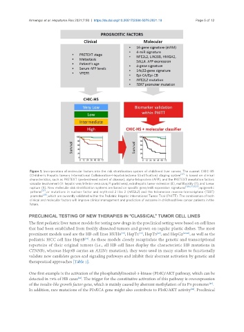
Figure 1. Incorporation of molecular factors into the risk stratification system of childhood liver cancer. The current CHIC-HS
[37]
(Children’s Hepatic tumors International Collaboration-Hepatoblastoma Stratification) staging system is based on clinical
characteristics, such as PRETEXT (pretreatment extent of disease), alpha-fetoprotein (AFP), and the PRETEXT annotation factors
vascular involvement (V: hepatic vein/inferior vena cava; P: portal vein), extrahepatic tumor extension (E), multifocality (F), and tumor
[11,16,27,31,32]
rupture (R). New molecular risk stratification systems are based on specific gene/miR expression signatures , epigenetic
[27]
patterns , or mutations in nuclear factor and erythroid 2 like 2 (NFE2L2) and the telomerase reverse-transcriptase (TERT)
[26]
promoter , which are currently validated within the Pediatric Hepatic International Tumor Trial (PHITT). The combination of both
clinical and molecular factors will improve clinical management and prediction of outcome in childhood liver cancer patients in the
future.
PRECLINICAL TESTING OF NEW THERAPIES IN “CLASSICAL” TUMOR CELL LINES
The first pediatric liver tumor models for testing new drugs in the preclinical setting were based on cell lines
that had been established from freshly dissected tumors and grown on regular plastic dishes. The most
[39]
[38]
prominent models used are the HB cell lines HUH6 , HepT1 , HepT3 , and HepG2 [41,42] , as well as the
[40]
pediatric HCC cell line Hep3B . As these models closely recapitulate the genetic and transcriptional
[43]
repertoire of their original tumors (i.e., all HB cell lines display the characteristic HB mutations in
CTNNB1, whereas Hep3B carries an AXIN1 mutation), they were used in many studies to functionally
validate new candidate genes and signaling pathways and inhibit their aberrant activation by genetic and
therapeutical approaches [Table 1].
One first example is the activation of the phosphatidylinositol-3-kinase (PI3K)/AKT pathway, which can be
detected in 79% of HB cases . The trigger for the constitutive activation of this pathway is overexpression
[44]
of the insulin-like growth factor gene, which is mainly caused by aberrant methylation of its P3 promoter .
[45]
[44]
In addition, rare mutations of the PI3KCA gene might also contribute to PI3K/AKT activity . Preclinical