
Page 11 - Read Online
P. 11
Page 4 of 12 Hiyama et al. Hepatoma Res 2021;7:44 https://dx.doi.org/10.20517/2394-5079.2021.21
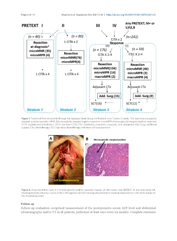
Figure 1. Treatment flow of patients through the Japanese Study Group for Pediatric Liver Tumors-2 study. *Six were macroscopically
marginal positive resection. MNR: Microscopically marginal negative resection; microMPR: microscopically marginal positive resection;
CITA: cisplatin and pirarubicin; L-CITA: low-dose CITA; ITEC: ifosfamide, pirarubicin, etoposide, and carboplatin; Add. Surg.: additional
surgery; CTx: chemotherapy; SCT: high-dose chemotherapy with stem cell transplantation.
Figure 2. A representative case of a microscopically positive resection margin: (A) this tumor was PRETEXT III and underwent left
hemihepatectomy after four cycles of the CITA regimen; and (B) histological examination revealed residual tumor cells at the margin of
the resected specimen.
Follow up
Follow-up evaluation comprised measurement of the postoperative serum AFP level and abdominal
ultrasonography and/or CT in all patients, performed at least once every six months. Complete remission