
Page 15 - Read Online
P. 15
Igarashi et al. Hepatoma Res 2022;8:21 https://dx.doi.org/10.20517/2394-5079.2022.02 Page 3 of 6
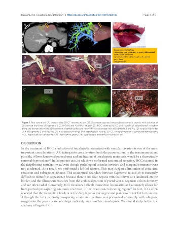
Figure 1. First operation: (A) preoperative 3D CT reconstruction; (B) Glissonean approach respecting Laennec’s capsule with isolation of
Glissonean branches of Segments 3 (G3) (left) and 4a (G4a) (right); (C) HCC staining by ICG and superficial parenchymal resection
along the demarcation line; (D) isolation of umbilical fissure vein (UFV) as drainage vein of Segments 3 and 4a; (E) surgical field after
LAR of Segments 3 and 4a; and (F) macroscopic findings and pathological results. 3D CT, three-dimensional computed tomography;
HCC, hepatocellular carcinoma; ICG, Indocyanine green; LAR, laparoscopic anatomical liver resection.
DISCUSSION
In the treatment of HCC, eradication of intrahepatic metastasis with vascular invasion is one of the most
important considerations. AR, taking into consideration both the preservation, to the maximum extent
possible, of liver functional parenchyma and eradication of intrahepatic metastasis, would be a theoretically
[1]
reasonable procedure . In the present case, in which we performed anatomical resection, HCC recurred in
the neighboring segment twice, even though pathological vascular invasion and marginal remnants were
not confirmed. As a result, we performed a left lobectomy. This may suggest a limitation of cone unit
resection and subsegmentectomy. The anatomical boundary between Segments 4a and 4b is extremely
difficult to identify in appearance because there is no clear hepatic vein that serves as a landmark on the
border, and the Glissonean branches from the umbilical portion of portal vein to Segment 4 show diversity
and are often radial. Conversely, ICG visualizes difficult transection boundaries and ultimately allows for
liver parenchyma-sparing anatomic resection of the exact cancer-bearing region . In fact, ICG often
[9]
revealed that the transection borders in the deep layer as intersegmental planes were not flat but uneven.
Although the liver parenchyma-sparing anatomic resection was performed accurately with adequate
margins for the present case, oncologic radicality may have been inadequate. We should study further the
anatomy of Segment 4.