
Page 78 - Read Online
P. 78
Osho et al. Hepatoma Res 2020;6:55 I http://dx.doi.org/10.20517/2394-5079.2020.42 Page 7 of 15
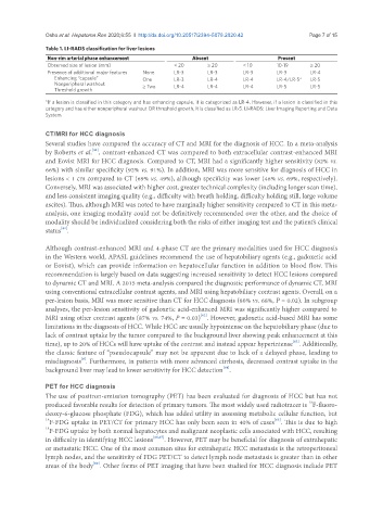
Table 1. LI-RADS classification for liver lesions
Non-rim arterial phase enhancement Absent Present
Observed size of lesion (mm) < 20 ≥ 20 < 10 10-19 ≥ 20
Presence of additional major features None LR-3 LR-3 LR-3 LR-3 LR-4
Enhancing “capsule” One LR-3 LR-4 LR-4 LR-4/LR-5* LR-5
Nonperipheral washout ≥ Two LR-4 LR-4 LR-4 LR-5 LR-5
Threshold growth
*If a lesion is classified in this category and has enhancing capsule, it is categorized as LR-4. However, if a lesion is classified in this
category and has either nonperipheral washout OR threshold growth, it is classified as LR-5. LI-RADS: Liver Imaging Reporting and Data
System
CT/MRI for HCC diagnosis
Several studies have compared the accuracy of CT and MRI for the diagnosis of HCC. In a meta-analysis
[61]
by Roberts et al. , contrast-enhanced CT was compared to both extracellular contrast-enhanced MRI
and Eovist MRI for HCC diagnosis. Compared to CT, MRI had a significantly higher sensitivity (82% vs.
66%) with similar specificity (92% vs. 91%). In addition, MRI was more sensitive for diagnosis of HCC in
lesions < 1 cm compared to CT (69% vs. 49%), although specificity was lower (46% vs. 69%, respectively).
Conversely, MRI was associated with higher cost, greater technical complexity (including longer scan time),
and less consistent imaging quality (e.g., difficulty with breath holding, difficulty holding still, large volume
ascites). Thus, although MRI was noted to have marginally higher sensitivity compared to CT in this meta-
analysis, one imaging modality could not be definitively recommended over the other, and the choice of
modality should be individualized considering both the risks of either imaging test and the patient’s clinical
[61]
status .
Although contrast-enhanced MRI and 4-phase CT are the primary modalities used for HCC diagnosis
in the Western world, APASL guidelines recommend the use of hepatobiliary agents (e.g., gadoxetic acid
or Eovist), which can provide information on hepatocellular function in addition to blood flow. This
recommendation is largely based on data suggesting increased sensitivity to detect HCC lesions compared
to dynamic CT and MRI. A 2015 meta-analysis compared the diagnostic performance of dynamic CT, MRI
using conventional extracellular contrast agents, and MRI using hepatobiliary contrast agents. Overall, on a
per-lesion basis, MRI was more sensitive than CT for HCC diagnosis (80% vs. 68%, P = 0.02). In subgroup
analyses, the per-lesion sensitivity of gadoxetic acid-enhanced MRI was significantly higher compared to
[62]
MRI using other contrast agents (87% vs. 74%, P = 0.03) . However, gadoxetic acid-based MRI has some
limitations in the diagnosis of HCC. While HCC are usually hypointense on the hepatobiliary phase (due to
lack of contrast uptake by the tumor compared to the background liver showing peak enhancement at this
[63]
time), up to 20% of HCCs will have uptake of the contrast and instead appear hyperintense . Additionally,
the classic feature of “pseudocapsule” may not be apparent due to lack of a delayed phase, leading to
[6]
misdiagnosis . Furthermore, in patients with more advanced cirrhosis, decreased contrast uptake in the
[64]
background liver may lead to lower sensitivity for HCC detection .
PET for HCC diagnosis
The use of positron-emission tomography (PET) has been evaluated for diagnosis of HCC but has not
18
produced favorable results for detection of primary tumors. The most widely used radiotracer is F-fluoro-
deoxy-6-glucose phosphate (FDG), which has added utility in assessing metabolic cellular function, but
[65]
18 F-FDG uptake in PET/CT for primary HCC has only been seen in 40% of cases . This is due to high
18 F-FDG uptake by both normal hepatocytes and malignant neoplastic cells associated with HCC, resulting
in difficulty in identifying HCC lesions [66,67] . However, PET may be beneficial for diagnosis of extrahepatic
or metastatic HCC. One of the most common sites for extrahepatic HCC metastasis is the retroperitoneal
lymph nodes, and the sensitivity of FDG PET/CT to detect lymph node metastasis is greater than in other
[68]
areas of the body . Other forms of PET imaging that have been studied for HCC diagnosis include PET