
Page 66 - Read Online
P. 66
Vezeridis et al. Hepatoma Res 2020;6:53 I http://dx.doi.org/10.20517/2394-5079.2020.36 Page 5 of 10
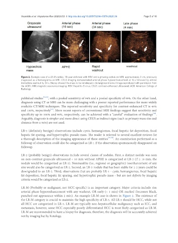
Figure 2. Example case of a LR-M nodule. 78-year-old male with HBV and a growing nodule on MRI, approximately 3 cm, previously
diagnosed as a hemangioma on MRI. CEUS imaging demonstrated arterial phase hyperenhancement at 16 s followed by almost
immediate washout by 34 s. Biopsy showed the mass to be intrahepatic cholangiocarcinoma (image reproduced with permission from
the ACR). MRI: magnetic resonance imaging; HBV: hepatitis B virus; CEUS: contrast-enhanced ultrasound; ACR: American College of
Radiology
published studies [31,32] , with a pooled sensitivity of 94% and a pooled specificity of 99%. On the other hand,
diagnosis using CT or MRI can be more challenging with a poorer reported performance for more widely
available CT/MRI techniques. The reported sensitivity and specificity for contrast-enhanced CT is 43%
[33]
and 100%, respectively . More recent reports of conventional MRI findings suggest that sensitivity and
[34]
specificity up to 100% and 90%, respectively, can be achieved with a “careful” evaluation of findings .
Arguably, diagnosis is simpler and more direct using CEUS as indirect signs (such as primary mass size and
distance from a vein) are not used.
LR-1 (definitely benign) observations include cysts, hemangiomas, focal hepatic fat deposition, focal
hepatic fat sparing, and hypertrophic pseudo mass. The reader is referred to several excellent reviews for
a thorough description of the imaging appearance of these entities [35-39] . An examination performed as a
followup of observation could also be categorized as LR-1 if the observation spontaneously disappeared on
followup.
LR-2 (probably benign) observations include several classes of nodules. First, a distinct nodule was seen
on non-contrast grayscale ultrasound < 10 mm without APHE is categorized at LR-2 (if ≥ 10 mm, the
nodule would be categorized as LR-3). Nonmasslike (i.e., regional or geographic) isoenhancement of any
size would also be categorized as LR-2. Second, an LR-3 nodule that has been stable for ≥ 2 years could be
downgraded to an LR-2. Third, observations that are probably LR-1 - cysts, hemangiomas, focal hepatic
fat deposition, focal hepatic fat sparing, and hypertrophic pseudo mass - but are not definite by imaging
criteria would be categorized as LR-2.
LR-M (Probably or malignant, not HCC specific) is an important category. Major criteria include rim
arterial phase hyperenhancement with any washout, OR early (< 1 min) OR marked (becomes black,
punched out appearance within 2 min). An example LR-M case is shown in Figure 2. The existence of
the LR-M category is crucial to maintain the high specificity of LR-5. All LR-5 should be HCC, while not
all HCC are categorized in LR-5. LR-M are typically non-hepatocellular malignancy such as ICC, and
metastasis, however, some HCC especially poorly differentiated HCC is most likely categorized as LR-M.
LR-M are recommended to have a biopsy for diagnosis, therefore, the diagnosis will be accurately achieved
not by imaging but by histology.