
Page 11 - Read Online
P. 11
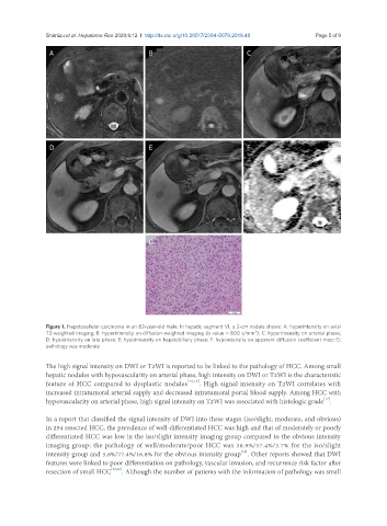
Shimizu et al. Hepatoma Res 2020;6:12  I  http://dx.doi.org/10.20517/2394-5079.2019.48                                              Page 5 of 9

                A                              B                             C


                D                              E                             F


                                               G


               Figure 1. Hepatocellular carcinoma in an 82-year-old male. In hepatic segment VI, a 2-cm nodule shows: A: hyperintensity on axial
                                                                               2
               T2-weighted imaging; B: hyperintensity on diffusion-weighted imaging (b value = 800 s/mm ); C: hyperintensity on arterial phase;
               D: hypointensity on late phase; E: hypointensity on hepatobiliary phase; F: hypointensity on apparent diffusion coefficient map; G:
               pathology was moderate

               The high signal intensity on DWI or T2WI is reported to be linked to the pathology of HCC. Among small
               hepatic nodules with hypovascularity on arterial phase, high intensity on DWI or T2WI is the characteristic
               feature of HCC compared to dysplastic nodules [15,16] . High signal intensity on T2WI correlates with
               increased intratumoral arterial supply and decreased intratumoral portal blood supply. Among HCC with
                                                                                                     [17]
               hypovascularity on arterial phase, high signal intensity on T2WI was associated with histologic grade .
               In a report that classified the signal intensity of DWI into three stages (iso/slight, moderate, and obvious)
               in 254 resected HCC, the prevalence of well-differentiated HCC was high and that of moderately or poorly
               differentiated HCC was low in the iso/slight intensity imaging group compared to the obvious intensity
               imaging group: the pathology of well/moderate/poor HCC was 38.9%/57.4%/3.7% for the iso/slight
                                                                           [18]
               intensity group and 5.8%/77.4%/16.8% for the obvious intensity group . Other reports showed that DWI
               features were linked to poor differentiation on pathology, vascular invasion, and recurrence risk factor after
               resection of small HCC [19,20] . Although the number of patients with the information of pathology was small