
Page 10 - Read Online
P. 10
Page 4 of 9 Shimizu et al. Hepatoma Res 2020;6:12 I http://dx.doi.org/10.20517/2394-5079.2019.48
Table 1. Patients’ characteristics
DAA-SVR HCC (n = 26) HCV-positive HCC (n = 80) P value
Age (years), median (range) 75 (63-83) 75 (44-93) 0.27
Sex (male/female) 10/16 43/37 0.26
Number of tumors (single/multiple) 21/5 60/20 0.61
Tumor diameter (cm), median (range) 18.0 (10-29) 17.6 (8.0-29) 0.7
BCLC stage A/B/C 26/0/0 80/0/0 1.0
AFP (ng/mL) 3.4 (2.0-6116.5) 25.3 (2.0-1660) < 0.001
PIVKA-II (mAU/mL) 19.0 (12-94) 53.0 (13-245) 0.2
Albumin (g/dL) 4.0 (2.9-4.7) 3.7 (2.8-273) 0.6
ALT (IU/mL) 18.0 (12-94) 53 (13-245) < 0.001
3
Platelet (10 /μL) 149 (45-189) 102 (33-344) 0.009
Total bilirubin (mg/dL) 0.7 (0.3-2.0) 0.8 (0.3-2.6) 0.71
PT% 91 (57-113) 89 (31-120) 0.24
Child-Pugh score 5/6 25/1 68/12 0.18
Fib 4 index 3.22(0.55-11.5) 6.01(1.66-25.2) < 0.001
BCLC: barcelona clinic liver cancer; AFP: alpha-fetoprotein; PIVKA-II: protein induced by vitamin K absence or antagonist-II; ALT: alanine
aminotransferase; PT: prothrombin time; DAA: direct-acting antiviral; SVR: sustained viral response; HCC: hepatocellular carcinoma;
HCV: hepatitis C virus
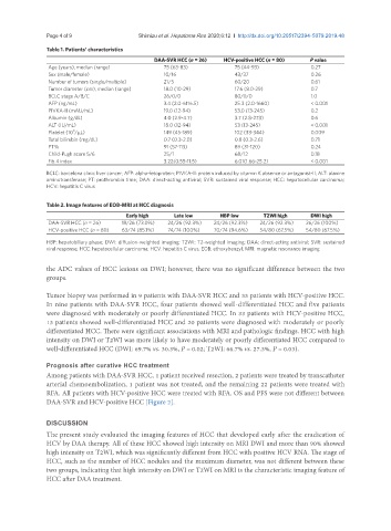
Table 2. Image features of EOB-MRI at HCC diagnosis
Early high Late low HBP low T2WI high DWI high
DAA-SVR HCC (n = 26) 19/26 (73.0%) 24/26 (92.3%) 24/26 (92.3%) 24/26 (92.3%) 26/26 (100%)
HCV-positive HCC (n = 80) 63/74 (85.1%) 74/74 (100%) 70/74 (94.6%) 54/80 (67.5%) 54/80 (67.5%)
HBP: hepatobiliary phase; DWI: diffusion-weighted imaging; T2WI: T2-weighted imaging; DAA: direct-acting antiviral; SVR: sustained
viral response; HCC: hepatocellular carcinoma; HCV: hepatitis C virus; EOB: ethoxybenzyl; MRI: magnetic resonance imaging
the ADC values of HCC lesions on DWI; however, there was no significant difference between the two
groups.
Tumor biopsy was performed in 9 patients with DAA-SVR HCC and 33 patients with HCV-positive HCC.
In nine patients with DAA-SVR HCC, four patients showed well-differentiated HCC and five patients
were diagnosed with moderately or poorly differentiated HCC. In 33 patients with HCV-positive HCC,
13 patients showed well-differentiated HCC and 20 patients were diagnosed with moderately or poorly
differentiated HCC. There were significant associations with MRI and pathologic findings. HCC with high
intensity on DWI or T2WI was more likely to have moderately or poorly differentiated HCC compared to
well-differentiated HCC (DWI: 69.7% vs. 30.3%, P = 0.02; T2WI: 66.7% vs. 27.3%, P = 0.03).
Prognosis after curative HCC treatment
Among patients with DAA-SVR HCC, 1 patient received resection, 2 patients were treated by transcatheter
arterial chemoembolization, 1 patient was not treated, and the remaining 22 patients were treated with
RFA. All patients with HCV-positive HCC were treated with RFA. OS and PFS were not different between
DAA-SVR and HCV-positive HCC [Figure 2].
DISCUSSION
The present study evaluated the imaging features of HCC that developed early after the eradication of
HCV by DAA therapy. All of these HCC showed high intensity on MRI DWI and more than 90% showed
high intensity on T2WI, which was significantly different from HCC with positive HCV RNA. The stage of
HCC, such as the number of HCC nodules and the maximum diameter, was not different between these
two groups, indicating that high intensity on DWI or T2WI on MRI is the characteristic imaging feature of
HCC after DAA treatment.