
Page 56 - Read Online
P. 56
Puppala. Hepatoma Res 2019;5:44 I http://dx.doi.org/10.20517/2394-5079.2019.28 Page 7 of 10
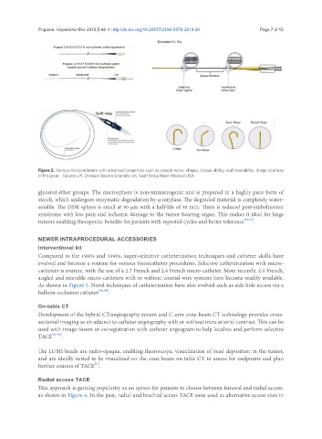
Figure 5. Various microcatheters with advanced properties such as coaxial wires, shapes, torque ability, and steerability. Image courtesy
of Pro great - Terumo UK, Direxion Boston Scientific UK, Swift Ninja Merit Medical USA
glycerol ether groups. The microsphere is non-immunogenic and is prepared in a highly pure form of
starch, which undergoes enzymatic degradation by α-amylase. The degraded material is completely water-
soluble. The DSM sphere is small at 50 µm with a half-life of 35 min. There is reduced post-embolization
syndrome with less pain and ischemic damage to the tumor-bearing organ. This makes it ideal for large
tumors enabling therapeutic benefits for patients with repeated cycles and better tolerance [32,33] .
NEWER INTRAPROCEDURAL ACCESSORIES
Interventional kit
Compared to the 1980s and 1990s, super-selective catheterization techniques and catheter skills have
evolved and become a routine for various transcatheter procedures. Selective catheterization with micro-
catheters is routine, with the use of a 2.7 French and 2.4 French micro-catheter. More recently, 2.0 French,
angled and steerable micro-catheters with or without coaxial wire systems have become readily available.
As shown in Figure 5. Novel techniques of catheterization have also evolved such as side hole access via a
balloon occlusion catheter [34,35] .
On-table CT
Development of the hybrid CT/angiography system and C-arm cone-beam CT technology provides cross-
sectional imaging as an adjunct to catheter angiography with or without intra-arterial contrast. This can be
used with image fusion or co-registration with catheter angiogram to help localize and perform selective
TACE [36-38] .
The LUMI beads are radio-opaque, enabling fluoroscopic visualization of bead deposition in the tumor,
and are ideally suited to be visualized on the cone-beam on-table CT to assess for endpoints and plan
[31]
further courses of TACE .
Radial access TACE
This approach is gaining popularity as an option for patients to choose between femoral and radial access,
as shown in Figure 6. In the past, radial and brachial access TACE were used as alternative access sites in