
Page 55 - Read Online
P. 55
Page 6 of 10 Puppala. Hepatoma Res 2019;5:44 I http://dx.doi.org/10.20517/2394-5079.2019.28
A B C
D E F
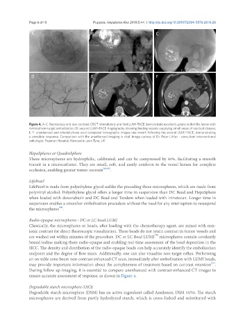
Figure 4. A-C: fluoroscopy and non-contrast CBCT immediately and first LUMI-TACE demonstrate excellent uptake within the lesion with
minimal non-target embolization; D: second LUMI-TACE Angiography showing feeding vessels supplying small areas of residual disease;
E, F: unenhanced and arterial-phase axial computed tomography images one month following the second LUMI-TACE, demonstrating
a complete response. Comparison with the unenhanced imaging is vital. Image curtesy of Dr. Peter Littler - consultant interventional
radiologist, Freeman Hospital, Newcastle upon Tyne, UK
HepaSpheres or QuadraSphere
These microspheres are hydrophilic, calibrated, and can be compressed by 80%, facilitating a smooth
transit in a microcatheter. They are small, soft, and easily conform to the vessel lumen for complete
occlusion, enabling greater tumor necrosis [28,29] .
LifePearl
LifePearl is made from polyethylene glycol unlike the preceding three microspheres, which are made from
polyvinyl alcohol. Polyethylene glycol offers a longer time in suspension than DC Bead and HepaSphere
when loaded with doxorubicin and DC Bead and Tandem when loaded with irinotecan. Longer time in
suspension enables a smoother embolization procedure without the need for any interruption to resuspend
[30]
the microspheres .
Radio-opaque microspheres - DC or LC bead LUMI
Classically, the microspheres or beads, after loading with the chemotherapy agent, are mixed with non-
ionic contrast for direct fluoroscopic visualization. These beads do not retain contrast in tumor vessels and
TM
are washed out within minutes of the procedure. DC or LC Bead LUMI microspheres contain covalently
bound iodine making them radio-opaque and enabling real-time assessment of the bead deposition in the
HCC. The density and distribution of the radio-opaque beads can help accurately identify the embolization
endpoint and the degree of flow stasis. Additionally, one can also visualize non-target reflux. Performing
an on-table cone-beam non-contrast enhanced CT scan, immediately after embolization with LUMI beads,
may provide important information about the completeness of treatment based on contrast retention .
[31]
During follow up imaging, it is essential to compare unenhanced with contrast-enhanced CT images to
ensure accurate assessment of response, as shown in Figure 4.
Degradable starch microsphere-TACE
Degradable starch microsphere (DSM) has an active ingredient called Amilomer, DSM 35/50. The starch
microspheres are derived from partly hydrolyzed starch, which is cross-linked and substituted with