
Page 49 - Read Online
P. 49
Page 4 of 10 Reyngold et al. Hepatoma Res 2018;4:49 I http://dx.doi.org/10.20517/2394-5079.2018.84
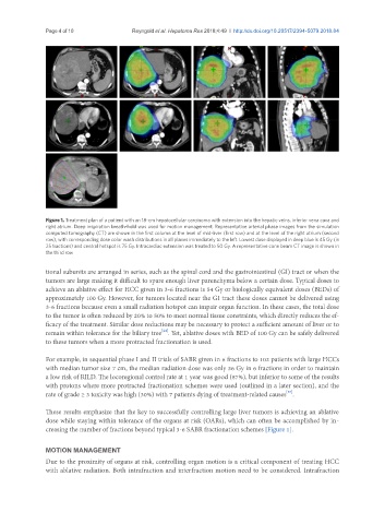
Figure 1. Treatment plan of a patient with an 18-cm hepatocellular carcinoma with extension into the hepatic veins, inferior vena cava and
right atrium. Deep inspiration breath-hold was used for motion management. Representative arterial phase images from the simulation
computed tomography (CT) are shown in the first column at the level of mid-liver (first row) and at the level of the right atrium (second
row), with corresponding dose color wash distributions in all planes immediately to the left. Lowest dose displayed in deep blue is 45 Gy (in
25 fractions) and central hotspot is 75 Gy. Intracardiac extension was treated to 50 Gy. A representative cone beam CT image is shown in
the third row
tional subunits are arranged in series, such as the spinal cord and the gastrointestinal (GI) tract or when the
tumors are large making it difficult to spare enough liver parenchyma below a certain dose. Typical doses to
achieve an ablative effect for HCC given in 3-6 fractions is 54 Gy or biologically equivalent doses (BEDs) of
approximately 100 Gy. However, for tumors located near the GI tract these doses cannot be delivered using
3-6 fractions because even a small radiation hotspot can impair organ function. In these cases, the total dose
to the tumor is often reduced by 20% to 50% to meet normal tissue constraints, which directly reduces the ef-
ficacy of the treatment. Similar dose reductions may be necessary to protect a sufficient amount of liver or to
[24]
remain within tolerance for the biliary tree . Yet, ablative doses with BED of 100 Gy can be safely delivered
to these tumors when a more protracted fractionation is used.
For example, in sequential phase I and II trials of SABR given in 6 fractions to 102 patients with large HCCs
with median tumor size 7 cm, the median radiation dose was only 36 Gy in 6 fractions in order to maintain
a low risk of RILD. The locoregional control rate at 1 year was good (87%), but inferior to some of the results
with protons where more protracted fractionation schemes were used (outlined in a later section), and the
[25]
rate of grade ≥ 3 toxicity was high (30%) with 7 patients dying of treatment-related causes .
These results emphasize that the key to successfully controlling large liver tumors is achieving an ablative
dose while staying within tolerance of the organs at risk (OARs), which can often be accomplished by in-
creasing the number of fractions beyond typical 3-6 SABR fractionation schemes [Figure 1].
MOTION MANAGEMENT
Due to the proximity of organs at risk, controlling organ motion is a critical component of treating HCC
with ablative radiation. Both intrafraction and interfraction motion need to be considered. Intrafraction