Page 179 - Read Online
P. 179
Paranaguá-Vezozzo et al. Hepatoma Res 2018;4:11  I  http://dx.doi.org/10.20517/2394-5079.2018.17                         Page 5 of 11


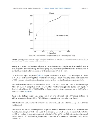
               Figure 1. Receiver operator curve analysis of calculated model score for identifying hepatocellular carcinoma. ALT: alanine
               aminotransferase; AFP: alpha-fetoprotein; AUC: area under curve

               Among HCC patients, 19 (61%) were subjected to antiviral treatment with alpha-interferon, to which none of
               them responded. However, among the control group, 42 (68%) were subjected to antiviral treatment, and 10
               (24%) of these patients achieved sustained virological response (SVR) (P = 0.05).

               On multivariate logistic regression [Table 2], higher AFP levels (> 20 ng/mL, P < 0.001), higher ALT levels
                                                                 3
               (> 37 U/L, P = 0.01) and lower platelet count (< 100,000/mm , P = 0.007) were independent prediction factors
               of HCC presence, with odds ratios of 16.2 (4.17-63.01), 7.43 (1.61-34.19) and 3.62 (1.43-9.14), respectively.


               The coefficients of the multivariable model are 3.71 ± 1, 2.96 ± 0.77, 1.72 ± 0.9, 1.7 ± 0.62 for the intercept,
               AFP > 20, ALT > 37 and platelet count < 100,000. These variables were applied to build a score capable of
               discriminating higher risk of HCC in HCV cirrhotic patients, with an area under curve (AUC) of 0.79
               (95% CI: 0.7-0.89) [Figure 1].

               Based on the findings, we propose a model score to apply to outpatients with HCV related cirrhosis, but
               without tumors or nodules on US or CT/MRI images undertaken during routine surveillance:

               HCC Risk Score in HCV patients with cirrhosis = 46 × (abnormal AFP) + 27 × (abnormal ALT) + 27 × (abnormal
               platelet count)

               This formula requires the knowledge of the range and limits of the normal values of the aforementioned
               variables. For example, if AFP > 20 ng/dL, it is considered abnormal, and the score attributable to this
               variable is 1 (1), but if it ≤ 20 ng/dL its score is 0. Similarly, if the ALT is > 37 U/L, it is considered abnormal,
                                                                      3
               and the score is 1 (1), and finally a platelet count < 100,000/mm is considered an abnormal value, and its
               score is 1 (1).

