
Page 20 - Read Online
P. 20
Naeem et al. Hepatoma Res 2018;4:18 I http://dx.doi.org/10.20517/2394-5079.2018.22 Page 3 of 10
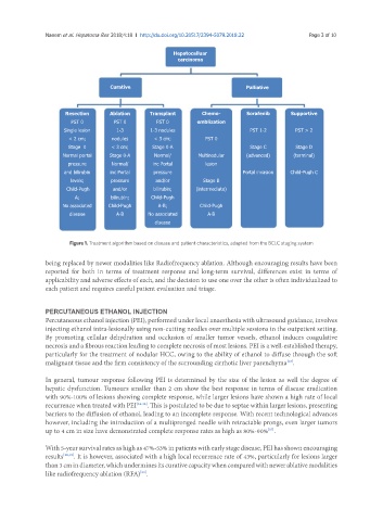
Hepatocelluar
carcinoma
Curative Palliative
Resection Ablation Transplant Chemo- Sorafenib Supportive
PST 0 PST 0 PST 0 emblization
Single lesion 1-3 1-3 nodules PST 1-2 PST > 2
< 2 cm; nodules < 3 cm; PST 0
Stage 0 < 3 cm; Stage 0-A Stage C Stage D
Normal portal Stage 0-A Normal/ Multinodular (advanced) (terminal)
pressure Normal/ inc Portal lesion
and bilirubin inc Portal pressure Portal invasion Child-Pugh C
levels; pressure and/or Stage B
Child-Pugh and/or bilirubin; (intermediate)
A; bilirubin; Child-Pugh
No associated Child-Pugh A-B; Child-Pugh
disease A-B No associated A-B
disease
Figure 1. Treatment algorithm based on disease and patient characteristics, adapted from the BCLC staging system
being replaced by newer modalities like Radiofrequency ablation. Although encouraging results have been
reported for both in terms of treatment response and long-term survival, differences exist in terms of
applicability and adverse effects of each, and the decision to use one over the other is often individualized to
each patient and requires careful patient evaluation and triage.
PERCUTANEOUS ETHANOL INJECTION
Percutaneous ethanol injection (PEI), performed under local anaesthesia with ultrasound guidance, involves
injecting ethanol intra-lesionally using non-cutting needles over multiple sessions in the outpatient setting.
By promoting cellular dehydration and occlusion of smaller tumor vessels, ethanol induces coagulative
necrosis and a fibrous reaction leading to complete necrosis of most lesions. PEI is a well-established therapy,
particularly for the treatment of nodular HCC, owing to the ability of ethanol to diffuse through the soft
malignant tissue and the firm consistency of the surrounding cirrhotic liver parenchyma .
[13]
In general, tumour response following PEI is determined by the size of the lesion as well the degree of
hepatic dysfunction. Tumours smaller than 2 cm show the best response in terms of disease eradication
with 90%-100% of lesions showing complete response, while larger lesions have shown a high rate of local
recurrence when treated with PEI [14-16] . This is postulated to be due to septae within larger lesions, presenting
barriers to the diffusion of ethanol, leading to an incomplete response. With recent technological advances
however, including the introduction of a multipronged needle with retractable prongs, even larger tumors
up to 4 cm in size have demonstrated complete response rates as high as 80%-90% .
[17]
With 5-year survival rates as high as 47%-53% in patients with early stage disease, PEI has shown encouraging
results [18,19] . It is however, associated with a high local recurrence rate of 43%, particularly for lesions larger
than 3 cm in diameter, which undermines its curative capacity when compared with newer ablative modalities
like radiofrequency ablation (RFA) .
[20]