
Page 208 - Read Online
P. 208
Page 4 of 6 Ohtsuka et al. Hepatoma Res 2024;10:18 https://dx.doi.org/10.20517/2394-5079.2024.04
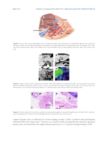
Figure 1. Course of the replaced left hepatic artery from the left gastric artery and direction of lymph flow. Blue arrows indicate the
direction of lymph flow from the left-sided liver and black arrows indicate the direction of lymph flow from the stomach. CEA: celiac
axis; CHA: common hepatic artery; SPA: splenic artery; LGA: left gastric artery; R-LHA: replaced left hepatic artery; IVC: inferior vena
cava.
Figure 2. Images of lymph node metastasis from the ICC in the left-sided liver. A metastatic lymph node (arrowhead) was present
along the replaced left hepatic artery (arrow). (A) ICC was located in the lateral segment (axial view); (B) (coronal view); (C)
18
Multidetector- row computed tomography image; (D) F-fluorodecxyglucose positron emission tomography image.
Figure 3. Photomicrograph of a lymphatic vessel and a lymph node around the replaced left hepatic artery (R-LHA). (A) A lymphatic
vessel containing cancer cells (arrow); (B) A lymph node with metastasized cancer cells (arrow).
common hepatic artery is sufficient for accurate staging, as only 3 of the 12 patients with gastrohepatic
LNM had LNM only in these sites . However, in our series, LNM only along the left embryonic (aberrant)
[17]
hepatic artery was identified as the single metastatic pathway in 6 of 11 patients with gastrohepatic LNM.