
Page 51 - Read Online
P. 51
Page 51 Racchetti et al. Extracell Vesicles Circ Nucleic Acids 2023;4:44-58 https://dx.doi.org/10.20517/evcna.2023.03
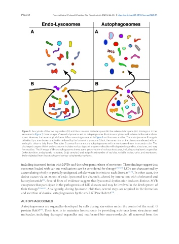
Figure 2. Exocytosis of the two organelles (B) and their released material spread in the extracellular space (A). Analogous to the
exosomes in Figure 1, these images of an endo-lysosome and an autophagosome illustrate exocytoses with release to the extracellular
space. However, the two exocytosis forms differ concerning exosomes in Figure 1 and from one another. The endo-lysosome B image is
delimited by a membrane combination induced by the fusion of a lysosome (black, the same color as the plasma membrane) with an
endocytic cisterna (sky blue). The other B comes from a mature autophagosome with a membrane drawn in a candy color. The
discharged cargoes (A) of endo-lysosome includes various types of enzyme molecules with degraded organelles, structures, and very
few vesicles. The A image of the autophagosome shows some preservation of various structures, including cytoplasmic organelles
(mitochondrion, endoplasmic reticulum, Golgi complex) and a significant number of vesicles, variable in size, color, and membrane,
likely originated from the autophagy of various cytoplasmic structures.
including increased fusion with MVBs and the subsequent release of exosomes. These findings suggest that
exosomes loaded with various medications can be considered for therapy [20,70,71] . LSDs are characterized by
accumulating wholly or partially undigested cellular waste intrinsic to each disorder [70-73] . In other cases, the
defect occurs via an excess of endo-lysosomal ion channels, altered by interaction with cholesterol and
lactosylceramide . Several lines of evidence suggest that lysosomal dysfunction induces distinct MVB
[70]
exocytoses that participate in the pathogenesis of LSD diseases and may be involved in the development of
their therapy [7,20,68,74] . Analogously, during lysosome inhibition, several steps are required in the formation
and secretion of classical autophagosomes by the small GTPase Rab27A .
[75]
AUTOPHAGOSOMES
Autophagosomes are organelles developed by cells during starvation under the control of the small G
[75]
protein Rab37 . Their task is to maintain homeostasis by providing nutrients from structures and
molecules, including damaged organelles and malformed bio-macromolecule, all removed from the