
Page 29 - Read Online
P. 29
Chakraborty et al. Extracell Vesicles Circ Nucleic Acids 2023;4:27-43 https://dx.doi.org/10.20517/evcna.2023.05 Page 29
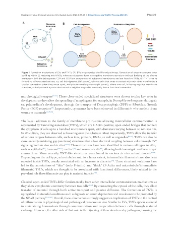
Figure 1. Formation mechanisms of EVs and TNTs. (A) EVs are generated via different pathways. Generation of exosomes require inner
budding within EE maturing into MVBs, whereas ectosomes form via negative membrane curvature-induced budding at the plasma
membrane. Both the tetraspanins CD9 and CD81 are components of ectosomal membranes and are found on TNTs. (B) TNTs can be
formed via different mechanisms, viz. cell dislodgement (left panels), wherein cells that come in contact with each other leave behind a
tubular connection when they move apart, and protrusion-elongation (right panels), where one cell, following negative membrane
curvature, actively extends a protrusion towards a neighbouring cell to eventually form a functional connection.
morphological synapses [21-23] . These close-ended specialized structures were shown to play key roles in
development as they allow the spreading of morphogens, for example, in Drosophila melanogaster during air
sac primordium’s development, through the transport of Decapentaplegic (DPP) or Fibroblast Growth
Factor (FGF) receptors . Importantly, cytonemes have been observed in different in vivo models, from
[24]
worms to mammals [22,25,26] .
The latest addition to the family of membrane protrusions allowing intercellular communication is
represented by Tunneling nanotubes (TNTs), which are F-Actin-positive, open-ended bridges that connect
the cytoplasm of cells up to a hundred micrometers apart, with diameters varying between 50 nm-900 nm.
In 2D culture, they are observed as hovering over the substrate. Most importantly, TNTs allow the transfer
of various cargoes between cells, such as ions, proteins, RNAs, as well as organelles [27-30] . TNTs can also be
2+
close-ended (containing gap junctions) structures that allow electrical coupling between cells through Ca
signaling both in vivo and in vitro [31-34] . These structures have been identified in various cell types in vitro,
[38]
[35]
such as epithelial , immune [36,37] , cardiac and neuronal cells , allowing both homotypic and heterotypic
[30]
connections. More recently TNT-like structures were found in various in vivo animal models [32,34] .
Depending on the cell type, microtubules and, to a lesser extent, intermediate filaments have also been
reported inside TNTs, usually associated with an increase in diameter . These structural variations have
[39]
led to the annotations of “thin” (only F-Actin) and “thick” (F-Actin and microtubule/intermediate
filaments) TNTs, which are thought to be associated with functional differences, likely related to the
prevalent role these filaments can play in material transfer .
[40]
Classical open-ended TNTs differ fundamentally from other intercellular communication mechanisms as
they allow cytoplasmic continuity between two cells [41,30] . By connecting the cytosol of the cells, they allow
transfer of material through both active transport and passive diffusion. The formation of TNTs is
upregulated in stressful conditions such as hypoxia or serum deprivation and was shown to be promoted by
the NF-κB pathway [32,42-44] . Overall, these observations strongly suggest an implication of TNTs in the control
of inflammation in physiological and pathological processes in vivo. Similar to EVs, TNTs appear essential
in maintaining homeostasis through communication and cooperation between cells through material
exchange. However, the other side of that coin is the hijacking of these structures by pathogens, favoring the