
Page 139 - Read Online
P. 139
Cai et al. Extracell Vesicles Circ Nucleic Acids 2023;4:262-82 https://dx.doi.org/10.20517/evcna.2023.10 Page 5
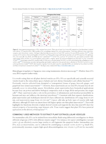
Figure 2. Heterogeneous populations of EVs isolated from plants. There are at least four known EV populations that have been isolated
from plants: TET-positive EVs, PEN1-positive EVs, autophagy-related EVs, and pollenosomes. They have different sizes, densities,
cargoes, and intracellular origins. Pathogen infection induces secretion of both TET-positive EVs and PEN1-positive EVs [56] . In the
process of EV isolation, final centrifugation of 40,000 × g (P40) pellets larger and heavier vesicles such as PEN1-positive EVs and large
EVs, non-vesicular free RNA, and RNA-protein complexes [30,79] . Small EVs, such as TET-positive EVs, are mainly present in the
supernatant after 40,000 × g centrifugation and require a higher speed of ultracentrifugation at 100,000 × g for collection
(P100) [30,56] . Autophagy-related EVs marked with ATG8a were collected using 100,000 × g from plants during the autophagy process
within cells [64] . Pollenosomes secreted during pollen germination and pollen tube growth were collected at 100,000 × g from in-vitro
pollen gemination media [59] . EXPO-derived EVs originate from the plant-specific organelle EXPO, are marked by the protein Exo70E2,
and have not yet been isolated from plants [71] . This figure was created with https://www.biorender.com/.
[55]
Rhizophagus irregularis or Gigaspora rosea using transmission electron microscopy . Whether these EVs
carry RNA requires further study.
It is worth noting that not all plant-derived vesicles are EVs. EVs are specifically and naturally secreted
vesicles found in the extracellular space of plants and have distinct biomarkers and cellular functions [30,45] .
Previously, vesicles collected from disrupted or homogenized tissues of grape, broccoli, ginger, and
grapefruit were termed nanovesicles [72,73] . However, these nanovesicles are not true EVs, because they do not
naturally occur in extracellular spaces. Nevertheless, plant nanovesicles have biomedical applications
because they can protect and deliver biological compounds, such as drugs, RNAs and proteins, into target
cells . Plant nanovesicles play a role in the prevention of inflammation and intestinal permeability in
[74]
[75]
humans and mice and influence the intestinal microbiome . Consistent with their separate classifications,
plant nanovesicles and Arabidopsis-derived EVs have different size distributions: EVs range from 60-200
nm, and nanovesicles range from 100-300 nm . Cancer cells take up both types of vesicles with high
[76]
efficiency, although EVs have an almost three-fold higher uptake rate than plant nanovesicles . This result
[76]
highlights the functional diversity of plant-derived vesicles and supports the idea that plant EVs have the
natural property of transporting cargo between cells. These results suggest that plant EVs have potential
applications in the delivery of therapeutics [77,78] .
COMMONLY USED METHODS TO EXTRACT PLANT EXTRACELLULAR VESICLES
For mammalian cells, EVs can be isolated from extracellular fluids using differential centrifugation to obtain
[9]
different subgroups of EVs with different density ranges . For instance, low-speed centrifugation (around
2,000 × g) can effectively recover large vesicles or cell fragments like apoptotic bodies. Intermediate-size
EVs, such as microvesicles, can be collected by centrifuging at around 10,000-20,000 × g. Small EVs,
predominantly exosomes, require high-speed ultracentrifugation over 100,000 × g (P100) for successful