
Page 56 - Read Online
P. 56
Page 166 Elton et al. Cancer Drug Resist 2020;3:161-70 I http://dx.doi.org/10.20517/cdr.2019.117
A B
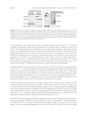
Figure 3. TOP2α/90 can be detected in both nuclear and cytoplasmic extracts and heterodimerizes with TOP2α/170. A: TOP2α
immunoassay using K562 and K/VP.5 cytoplasmic and nuclear lysates [36] . Immunoblots were probed with TOP2α/90/170 and β-actin
antibodies; B: immunoprecipitation experiments were performed using K562 and K/VP.5 whole cell lysates. The precipitated immune
complexes were released in SDS-PAGE sample buffer, subjected to SDS-PAGE, and immunoblotted, using the indicated antibodies. Input
immunoblots are also shown for each experiment and β-actin antibody loading controls. (A, B) Images are reproduced/adapted from
Figures 2B and 3D, respectively, published originally in Molecular Pharmacology; Kanagasabai et al. [36] , 2018. TOP2α: topoisomerase IIα;
SDS-PAGE: sodium dodecyl sulfate-polyacrylamide gel electrophoresis
It was hypothesized that, similar to the Top2α/160 truncated isoforms described above [32-34] , TOP2α/90
would be predominantly located in the cytoplasm since this isoform does not contain NLS 1454-1497
[Figure 1B]. Surprisingly, however, immunoassays using fractionated cytoplasmic and nuclear extracts
[Figure 3A] and immunofluorescence experiments (not shown) demonstrated that TOP2α/90 was
[36]
predominantly detected in the nucleus of K562 and K/VP.5 cells . Currently, it is not known how
TOP2α/90 is transported into the nucleus; a plausible speculation is that TOP2α/90 enters nuclei by a
[58]
“piggy-back” mechanism (e.g., heterodimerization) with TOP2α/170, since the full-length isoform
[47]
harbors functional NLS. In addition, TOP2α/90 may contain operative NLS sites. Mirski et al. found
three bipartite NLS sequences in the first 743 TOP2α aa but these were not functional. A short non-
[58]
classical IK-NLS motif (KVSKNK) in TOP2α/90 is currently under study for functionality.
Although TOP2α/90 does not harbor the DD essential for TOP2a/170:TOP2a/170 homodimerization
[Figure 1B] [41-45] , co-immunoprecipitation experiments demonstrated that endogenous TOP2α/90 and
[36]
TOP2α/170 proteins form heterodimers in both K562 and K/VP.5 cells [Figure 3B] . While these
results were unexpected, several studies have shown that human N-terminal TOP2α fragments, which
encompass just the ATPase domain (i.e., aa 1-435), dimerize in vitro under the appropriate conditions [59-61] .
[45]
Importantly, Bjergbaek et al. established that, if the C-terminal primary DD present in TOP2α/170 were
deleted, dimerization could still occur in the presence of DNA and an ATP analog.
Given that TOP2α/90 lacks the active site tyrosine residue (Tyr805) required to form TOP2α/170-DNA
covalent complexes [Figure 1B-iii], and is capable of heterodimerization with TOP2α/170 [Figure 3B],
it was posited that this isoform may be dominant-negative relative to drug-induced DNA damage and
cytotoxicity. Consistent with this hypothesis, forced overexpression of TOP2α/90 in K562 cells (which
express low levels of Top2α/90) decreased etoposide-induced DNA damage and cytotoxicity in K562
cells [Figure 4] [35,36] . Conversely, etoposide-induced DNA strand breaks were increased in K/VP.5 cells
subsequent to siRNA knockdown of elevated levels of TOP2α/90 [Figure 4] [35,36] .
Initial qPCR evaluation of paired AML patient samples (pre-treatment and relapse) indicated an increase in
[36]
the ratio of expression of TOP2α/90 mRNA compared to TOP2α/170 after relapse . In addition, the ratio
[36]
of TOP2α/90 to TOP2α/170 protein was increased after treatment relapse . To date, in four of six AML
patients, there was a statistically significant increase in the TOP2α/90 to TOP2α/170 ratio after relapse