
Page 22 - Read Online
P. 22
Page 342 Seno et al. Cancer Drug Resist 2019;2:335-50 I http://dx.doi.org/10.20517/cdr.2019.01
G H
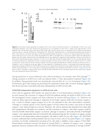
Figure 2. Daunorubicin induces apoptotic cell death in miPS-LLCcm cells with DNA fractionation. A: Morphology of miPS-LLCcm cells
undergoing cell death. Cells were treated with daunorubicin at a concentration of 100 nmol/L. Images were captured after 12 h of
treatment. Bar: 50 mm. Red arrows show dying miPS-LLCcm cells, and blue arrows show non-apoptotic cells; B: detection of apoptotic
cells by annexin-V staining. miPS-LLCcm were treated with 100 nmol/L daunorubicin for 3 h, harvested, and stained with annexin-V
APC. Flow cytometry was performed twice, and the representative results are shown; C: detection of oligonucleosomal DNA ladder in
bulk of miPS-LLCcm and in stem cells. DNA was prepared from miPS-LLCcm bulk (left) or puromycin-treated (right) cells subsequently
treated with 100 nmol/L daunorubicin for the indicated periods and subjected to agarose gel electrophoresis. Nuclear accumulation of
p53 protein following daunorubicin treatment; D: miPS-LLCcm cells were treated with daunorubicin for the indicated periods. Whole
cell lysates prepared and analyzed by western blotting using p53 antibody. β-actin levels were used as internal control; E: nuclear
accumulation of p53 proteins at the indicated periods was fractionated and subjected to western blotting. M: Mitochondrial, C:
cytoplasmic, and N: nuclear. β-tubulin and Histone H3 were detected for fractionation markers; F: expression of p53-regulated genes in
daunorubicin-treated miPS-LLCcm cells. RT-qPCR was performed using cDNA prepared from cells treated with 100 nmol/L daunorubicin
for the indicated periods (n = 3); G: suppression of Nanog gene expression was confirmed by RT-qPCR. Error bars in Figure 2E and F are
shown as SD; H: reduction in Nanog protein levels after daunorubicin treatment. Whole-cell lysates prepared from daunorubicin-treated
cells were subjected to western blot analysis
Nanog expression in mouse embryonic stem cells by binding to its promoter after DNA damage [21,22] .
Nanog expression in miPS-LLCcm cells was reduced within 3 h after daunorubicin treatment [Figure 2G].
In addition, Nanog protein levels were dramatically reduced after of 3 to 6 h of treatment [Figure 2H].
Furthermore, given that Nanog was predominantly expressed in stem cells, the above findings suggested
that the p53 pathway is activated in the stem cell population of miPS-LLCcm cells.
ICAD/CAD-independent apoptosis in miPS-LLCcm cells
Given that the apoptotic DNA ladder was observed after 12 h of daunorubicin treatment [Figure 2C],
we next assessed the activation of caspases-ICAD/CAD pathway during cell death of miPS-LLCcm cells.
We investigated several caspases and their substrates by western blotting [Figure 3A]. However, we
could not detect significant cleavage or reduction of full-length caspases [both the executioner (caspase-3
and -7) and the initiator caspase (caspase-9)] in the cells attached on dish after daunorubicin treatment.
Although an antibody specific to the cleaved caspase-9 failed to detect the protein, trace levels of cleaved
caspase-3 were observed (data not shown). Therefore, the cells in medium that detached from their dish
after programmed cell death were analyzed, and the activation of caspase 3 and other caspases could be
detected. Results showed that caspase-3 activation, cleavage of caspase-9, and reduced levels of full-length
caspase-7 were significantly detected in detached cells, demonstrating that the activation of caspase-cascade
is involved in the daunorubicin-induced apoptotic cell death of miPS-LLCcm cells [Figure 3B]. SDS-PAGE
analysis confirmed that overall proteolysis did not occur in these cells at this point [Figure 3C]. Caspase
activation was further confirmed by treatment of the cells with an inhibitor of pan-caspase, Z-VAD-FMK.
Daunorubicin treatment increased the levels of cleaved PARP-1, a substrate of caspase-3 and -7, during
apoptosis; however, treatment with 50 μmol/L Z-VAD-FMK blocked the accumulation of cleavage of PARP-