
Page 104 - Read Online
P. 104
Page 120 Gurule et al. Cancer Drug Resist 2018;1:118-25 I http://dx.doi.org/10.20517/cdr.2018.12
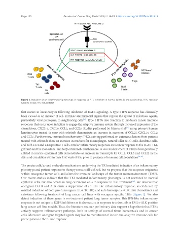
Figure 1. Induction of an inflammatory phenotype in response to RTK inhibition in normal epithelia and carcinomas. RTK: receptor
tyrosine kinase; NK: natural killer
that occurs in keratinocytes following inhibition of EGFR signaling. A type I IFN response has classically
been viewed as an inducer of cell- intrinsic antimicrobial signals that repress the spread of infectious agents,
particularly viral pathogens, to neighboring cells . Type I IFNs also function to modulate innate immune
[28]
responses that occur upon infection to engage the adaptive immune system through increased expression of the
chemokines, CXCL10, CXCL9, CCL2, and CCL5. Studies performed by Mascia et al. using primary human
[13]
keratinocytes treated in vitro with erlotinib demonstrate an increase in secretion of CCL27, CXCL14, CCL2
and CCL5. Furthermore, immunohistochemistry (IHC) staining performed on cutaneous lesions from patients
treated with erlotinib show an increase in markers for macrophages, natural killer (NK) cells, dendritic cells,
and both CD4 and CD8 positive T cells. Similar inflammatory responses are seen in response to the EGFR TKI,
gefitinib and the monoclonal antibody cetuximab. Furthermore, in vivo studies where EGFR has been genetically
ablated in murine epidermal cells demonstrates an increase in transcripts for CCL2, CCL5 and CCL22 in the
skin and circulation within their first week of life, prior to presence of immune cell populations [12,13] .
The precise cellular and molecular mechanisms underlying the TKI mediated induction of an inflammatory
phenotype and patient response to therapy remains ill-defined, but we propose that this response originates
within oncogenic tumor cells and alters the immune landscape of the tumor microenvironment (TME).
Our recent studies indicate that the TKI mediated inflammatory phenotype is not restricted to normal
epithelial cells, but also occurs in lung carcinoma cells in response to TKI treatment . We observe that
[34]
oncogenic EGFR and ALK cause a suppression of an IFN like inflammatory response, as evidenced by
marked induction of both pro-tumorigenic (IL6, TGFB2) and anti-tumorigenic (CXCL10) chemokines and
cytokines following treatment of lung cancer cell lines with oncogene specific TKIs [Figure 1]. We also
detect induction of these genes in on-treatment patient lung tumor samples. This IFN-like inflammatory
response is not unique to EGFR inhibitors as it also occurs in response to crizotinib in EML4-ALK positive
lung cancer cell line models. Thus, the literature and our preliminary data support a hypothesis that RTKs
actively suppress inflammatory pathways, both in settings of normal tissue homeostasis and in cancer
cells. Moreover, oncogene targeted agents may lead to recruitment of innate and adaptive immune cells for
participation in the tumor response.