
Page 14 - Read Online
P. 14
Page 616 Laubach et al. Cancer Drug Resist 2023;6:611-41 https://dx.doi.org/10.20517/cdr.2023.60
mounting evidence that accumulation of lactic acid within T cells dampens their function. Researchers have
+
also found that lactate, when studied separately from H in the form of sodium lactate, induces stemness
[45]
and tumor infiltration, and reduces apoptosis in CD8 T cells . Moreover, sodium lactate supplementation
+
in three mouse tumor models showed synergistic effects with anti-PD-1 treatment . A plausible
[45]
explanation for these somewhat contradictory findings is that variations between the TIMEs of different
tumor types metabolically reprogram CD8 TILs in distinct ways, wherein some tumors drive increased
+
sensitivity of CD8 TILs to lactic acid. Therefore, it is exceedingly important to delineate the metabolic
+
changes in CD8 TILs from different tumor types to identify the most effective therapy.
+
Additional research is needed to tease apart the intricate relationship between lactate, lactic acid, tumor
cells, CD8 T cells, and immunosuppressive cells. Inhibiting tumor-derived lactic acid production seems to
+
generally have anti-tumor effects, due to the detrimental effects of high acidity on the anti-tumor immune
cells within the TIME. While lactate ions serve as a carbon source and promote CD8 T cell stemness, they
+
also benefit immunosuppressive cells and excess amounts can dampen T cell effector functions. Collectively,
these data demonstrate that tumor-derived alterations in lactic acid metabolism contribute to ICB resistance
and modulating these pathways may augment efficacy, prompting the need for continued research efforts in
this field.
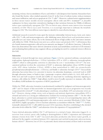
Adenosine
Adenosine is formed through two major pathways [Figure 1]. In the canonical pathway, ectonucleoside
triphosphate diphosphohydrolase-1 (CD39) hydrolyzes ATP or ADP to adenosine monophosphate
(AMP) , which is subsequently converted to adenosine by ecto-5′-nucleotidase (CD73) . The non-
[47]
[46]
canonical pathway involves the conversion of NAD to adenosine diphosphate ribose (ADPR) through
+
cyclic ADP ribose hydrolase (CD38); ADPR is then metabolized to AMP via ectonucleotide
pyrophosphatase/phosphodiesterase 1 (CD203a), and finally to adenosine through CD73 . Extracellular
[48]
adenosine has several fates; it is converted to inosine via adenosine deaminase, converted back to AMP
through adenosine kinase, or binds to type 1 purinergic receptors, which include A1, A2A, A2B, and A3.
Both A2A and A2B receptors (A2AR and A2BR) are important for mediating adenosine signaling in
immune cells within the TIME . High affinity A2AR is more broadly expressed on immune cells, while
[49]
[50]
low affinity A2BR facilitates the expansion of MDSC populations .
Within the TIME, adenosine formation is predominantly mediated by malignant and immunosuppressive
cells and the impact of this metabolite on immunosuppression and cancer progression was recently
[51]
[52]
comprehensively reviewed . Under physiological conditions, extracellular ATP and adenosine levels are
low . However, during cellular stress, such as hypoxia and nutrient deprivation, intracellular ATP is
[53]
released and serves as a strong pro-inflammatory mediator by recruiting immune cells [53,54] . On the other
hand, adenosine is a potent immunosuppressive metabolite . As such, it is not surprising that tumor cells
[50]
highly upregulate CD73 and immunosuppressive cells, such as cancer-associated fibroblasts (CAFs), Tregs,
and MDSCs, highly upregulate CD39 to facilitate adenosine accumulation within the TIME [52,55-59] . Further,
terminally exhausted CD8 T cells exhibit increased CD39 expression, therefore contributing to the elevated
+
adenosine levels within the TIME , and adenosine drives the expansion of Treg populations .
[61]
[60]
Tumor-derived adenosine inhibits CD8 T cell functions in a myriad of ways. Adenosine triggers IL-10
+
secretion from cervical cancer cells, leading to downregulation of MHC-I expression and subsequent
immune evasion from CD8 T cells . Increased adenosine production also favors tumor growth, as
+
[62]
indicated by the negative correlation between CD73 expression and survival in pancreatic adenocarcinoma
human cohorts . Moreover, loss of CD73 in pancreatic ductal adenocarcinoma cell lines leads to increased
[63]