
Page 135 - Read Online
P. 135
Spiliopoulou et al. Cancer Drug Resist 2024;7:2 https://dx.doi.org/10.20517/cdr.2023.46 Page 3
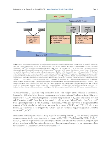
Figure 1. Main Mechanisms of Resistance (primary or secondary) to ICI. There are three different classifications or models summarizing
the main mechanisms of resistance to ICI. The first classification (blue triangles) describes the response to ICI in relationship to
markers of EMT [10] : the more tumors show a status of EMT, the lesser they respond to ICI. The second classification associates the
[8]
degree and type of fibrosis with responses to ICI (grey boxes) : response to ICI is generally observed in conditions with immune-
enriched fibrotic and non-fibrotic conditions. By contrast, immune-depleted or fibrotic conditions are not responsive to ICI. The third
classification is based on the presence of specific immune cells or markers (red boxes) [5,6] : responses to ICI are commonly observed in
+
patients with immune-inflamed conditions (characterized by a high CD8 /T cell ratio, B cells and TLS-rich tissues); conversely,
reg
responses are reduced in immune-excluded conditions (characterized by high vascular stroma content with fibrosis, chemokines, such
as CCL, CCL2, CCL5, CCL13, CCL22, or cytokines TGF-β). Limited or no responses to ICI are observed in patients with an immune-
deserted tumor microenvironment (lacking T cell priming, exhibiting tolerance, and displaying CAF-related markers). While T cells
reg
(green box) can be found in each of these conditions, their highest quantity and functional role are observed in either immune-excluded
conditions or in immune-enriched fibrotic tissues. ICI: Immune checkpoint inhibitor; EMT: epithelial-mesenchymal transition; TLS:
tertiary lymphoid structure; CCL: chemokine c-c-motif ligand; TGF-β: transforming growth factor beta; CAF: cancer-associated fibrosis.
“instructive model”, T cells are being “instructed” after T cell receptor (TCR) selection in the thymus.
Intermediate TCR stimulation (in contrast to negative and positive selection) leads to the intracellular gene
expression of FOXP3, which subsequently determines the generation of T cells. The second model is
reg
called “selection model”. According to this model, T cells are being “selected” rather than “instructed”
reg
from a pool of pre-formed T cells. According to this model, FOXP3 gene expression is independent of the
strength of TCR stimulation and further assumes the presence of FOXP3 and FOXP3 T cells in the
+
-
thymus. Upon exposure to self-antigens, the FOXP3 T cells are resistant to negative selection and form the
+
majority of T cells .
[25]
reg
Independent of the thymus, which is a key organ for the development of T cells, secondary lymphoid
reg
organs also appear to play a prominent role in generating CD4 FOXP3 T cells from CD4 FOXP3 T cells .
-
+
[26]
+
+
Such pT cells can originate from sub-immunogenic stimuli, non-inflammatory conditions, long-lasting or
reg
chronic infections, and inflammation. Furthermore, they are frequently present in various cancers where
they contribute to an immunosuppressive environment [27-30] .