
Page 68 - Read Online
P. 68
Stojkovska Docevska et al. Rare Dis Orphan Drugs J 2023;2:14 https://dx.doi.org/10.20517/rdodj.2023.09 Page 3 of 17
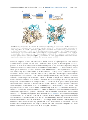
Figure 1. Structural characteristics of cathepsin C. (A) Schematic representation of the (pro)cathepsin C structure; (B) Schematic
representation of the active site of papain-like peptidases adapted for cathepsin C. The substrate binding sites on the enzyme are
labeled S3 through S2'. The substrate is presented with white circles corresponding to individual residues and residues P2 through P2'
are labelled. The arrow denotes the scissile bond; (C) Three-dimensional structure of a cathepsin C subunit. The peptidase domain is
shown in molecular surface representation and the exclusion domain in cartoon representation. Active site residues Cys234 and His381
are colored yellow and blue, respectively, Asp1 is shown as red sticks, and the chloride ion cofactor as a green sphere; (D) The
cathepsin C tetramer shown in cartoon representation. Exclusion domains are colored blue and peptidase domains are colored tan,
respectively. Coordinates were retrieved from the Protein Data Bank under accession code 1K3B.
removal of dipeptides from the N-terminus of the protein substrate. It stops only in three cases: when the
N-terminal amino group is blocked, when a proline residue is adjacent to the cleavage site (P1 or P1’
position), or when the N-terminal residues are lysine or arginine. Indeed, derivatives of positively charged
and aromatic amino acids have been found to competitively inhibit cathepsin C . Nevertheless, the enzyme
[20]
shows specific preferences for certain residues at positions P1 and P2 [16-18] and at positions P1’ and P2’ .
[19]
Due to its activity, most substrates used to measure cathepsin C activity in vitro are synthetic dipeptide
derivatives. The first reported substrates were Gly-Phe-p-nitroanilide (Gly-Phe-pNA) and Gly-Phe-β-
[15]
naphthylamide (Gly-Phe- βNA) . Their 7-amido-4-methylcoumarin analog (Gly-Phe-AMC) is the most
commonly used fluorogenic substrate in the published literature, although better substrates containing
natural and unnatural amino acids, such as L-norleucine, L-4-benzoylphenylalanine or L-glutamic acid
benzyl ester, have been identified by library screening [16-18] . In addition, highly specific internally quenched
substrates have been developed for the detection of cathepsin C activity in biological samples . Like most
[19]
PLPs, cathepsin C shows optimal activity under slightly acidic pH conditions [21,22] . Unlike others, it also
requires chloride (or other halides) ions for optimal activity below pH 7 [14,23] . At neutral and basic pH,
cathepsin C also exhibits transferase activity [24,25] , but similar activity has been observed with other related
and unrelated peptidases, e.g., papain . While most cysteine cathepsins are monomers, mature human
[26]
cathepsin C is a homotetramer organized as a dimer of dimers [13,22] [Figure 1D]. The exclusion domain plays
an indispensable role in tetramer formation, as each copy interacts with the peptidase domains of two
adjacent subunits. In the tetramer, the active sites of all subunits are fully exposed to the solvent, indicating
that all four subunits can function simultaneously. Interestingly, distant cathepsin C homologs have been
identified in unicellular eukaryotes, e.g., plasmodium, which were shown to be monomers . We have
[16]
recently conducted a phylogenetic and computational analysis of the evolution of cathepsin C and found
that the tetrameric form is likely restricted to animal cathepsin C homologs .
[4]