
Page 94 - Read Online
P. 94
Page 8 of 18 Kozarov et al. Vessel Plus 2020;4:10 I http://dx.doi.org/10.20517/2574-1209.2019.31
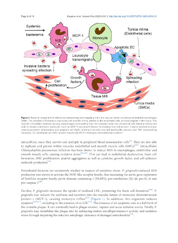
Figure 1. Bacterial component of atheromas representing transmigrating from the vascular lumen and tissue-embedded macrophages
(MΦ). The activated inflammatory leukocytes roll over the intima, adhere to the endothelial cells and transmigrate in the tissue. This
includes intracellular bacteria-carrying macrophages extravasating from the vascular lumen into the arterial wall. Bacteria induce host
cells to release chemotaxis molecules (such as MCP-1) and growth factors stimulating host cell division. Invasive bacteria multiply,
causing persistent inflammation and apoptotic cell death, forming a necrotic core and eroding the vascular wall. MN: extravasating
monocyte; EC: endothelial cell; SMC: smooth muscle cell; MCP-1: monocyte chemoattractant protein-1
[75]
intracellular, since they survive and multiply in peripheral blood mononuclear cells . They are also able
[105]
to replicate and persist within vascular endothelial and smooth muscle cells (SMCs) . Intracellular
Chlamydophila pneumoniae infection has been shown to induce ROS in macrophages, endothelial and
smooth muscle cells, causing oxidative stress [106,107] . This can lead to endothelial dysfunction, foam cell
formation, SMC proliferation, platelet aggregation as well as cytokine, growth factor, and cell adhesion
molecule production .
[101]
Periodontal bacteria are extensively studied in respect of oxidative stress. P. gingivalis-induced ROS
production was shown to activate the NOD-like receptor family, thus increasing the aortic gene expression
of Nod-like receptor family, pyrin domain containing 3 (NLRP3), pro-interleukin (IL)-1β, pro-IL-18 and
[108]
pro-caspase-1 .
Further, P. gingivalis increases the uptake of oxidized LDL, promoting the foam cell formation [109] . P.
gingivalis also induces the synthesis and secretion into the vascular lumen of monocyte chemoattractant
protein-1 (MCP-1), causing monocyte influx [110] [Figure 1]. In addition, this organism induces
apoptosis [91,92,111] , including in the presence of ox-LDL . The presence of an apoptotic core is a hallmark of
[112]
the unstable plaque. It can eventually lead to plaque erosion, rupture and acute ischemic events. Finally, P.
gingivalis may destabilize the plaque also by enhancing matrix metalloproteinase-9 activity and oxidative
[113]
stress through impairing the selective autophagic clearance of damaged mitochondria .