
Page 404 - Read Online
P. 404
Bader et al. Vessel Plus 2020;4:34 I http://dx.doi.org/10.20517/2574-1209.2020.36 Page 9 of 14
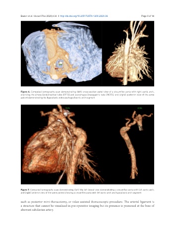
Figure 6. Computed tomography scan demonstrating (left) cross-section aerial view of a circumflex aorta with right aortic arch,
encircling the airway [endotracheal tube (ETT)] and oesophagus [nasogastric tube (NGT)]; and (right) posterior view of the same
patient demonstrating the hypoplastic retroesophageal aortic arch segment
Figure 7. Computed tomography scan demonstrating (left) the left lateral view demonstrating a circumflex aorta with left aortic arch;
and (right) anterior view of the same patient showing a circumflex aorta with left aortic arch and hypoplastic arch segment
such as posterior mini-thoracotomy, or video assisted thoracoscopic procedure. The arterial ligament is
a structure that cannot be visualized in pre-operative imaging but its presence is presumed at the base of
aberrant subclavian artery.