
Page 32 - Read Online
P. 32
Page 20 of 24 Rao. Vessel Plus 2022;6:22 https://dx.doi.org/10.20517/2574-1209.2021.105
Table 9. Causes of continuous murmurs. Modified from Rao [5]
Common causes
Patent ductus arteriosus
Venous hum
Surgical aorto-pulmonary shunts
Less common causes
Aorto pulmonary window
Persistent truncus arteriosus
Hemi truncus
Embryonic collateral vessels in pulmonary atresia with ventricular septal defect
Coronary arteriovenous fistula
Ruptured sinus of Valsalva aneurysm
Pulmonary arteriovenous fistula
Peripheral pulmonary artery stenosis
Coarctation of the aorta
Obstructed venous return
Cervical arteriovenous malformation
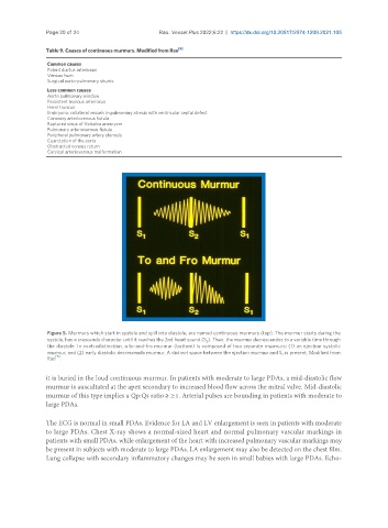
Figure 5. Murmurs which start in systole and spill into diastole, are named continuous murmurs (top). The murmur starts during the
systole, has a crescendo character until it reaches the 2nd heart sound (S ). Then, the murmur decrescendos to a variable time through
2
the diastole. In contradistinction, a to-and-fro murmur (bottom) is composed of two separate murmurs: (1) an ejection systolic
murmur; and (2) early diastolic decrescendo murmur. A distinct space between the ejection murmur and S is present. Modified from
2
[5]
Rao .
it is buried in the loud continuous murmur. In patients with moderate to large PDAs, a mid-diastolic flow
murmur is auscultated at the apex secondary to increased blood flow across the mitral valve. Mid-diastolic
murmur of this type implies a Qp:Qs ratio ≥ 2:1. Arterial pulses are bounding in patients with moderate to
large PDAs.
The ECG is normal in small PDAs. Evidence for LA and LV enlargement is seen in patients with moderate
to large PDAs. Chest X-ray shows a normal-sized heart and normal pulmonary vascular markings in
patients with small PDAs, while enlargement of the heart with increased pulmonary vascular markings may
be present in subjects with moderate to large PDAs. LA enlargement may also be detected on the chest film.
Lung collapse with secondary inflammatory changes may be seen in small babies with large PDAs. Echo-