


Page 66 - Read Online
P. 66
Bader et al. Vessel Plus 2020;4:34  I  http://dx.doi.org/10.20517/2574-1209.2020.36                                                  Page 5 of 14


                  A                                               B


                  C                                                   D


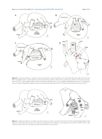
               Figure 3. Comparison between A: circumflex aorta with right aortic arch; B: double aortic arch with dominant right aortic arch (dominant
               RAA) with right common carotid (RCCA) right subclavian artery (RSCA), left common carotid artery (LCCA) and the left subclavian
               artery (LSCA); C: post-surgical division of the non-dominant distal left aortic arch in a patient with double aortic arch; D: double aortic
               arch with atretic left segment marked in red with the aberrant left subclavian (ALSCA) arising from Kommerell’s diverticulum (KD)


               Figure 4. Comparison between circumflex right aortic arch and common variant of right aortic arch with retroesophageal aberrant left
               subclavian artery (ALSCA) arising from Kommerell diverticulum (KD). Head and neck vessels are left common carotid (LCCA), right
               common carotid artery (RCCA), right subclavian artery (RSCA), and the ALSCA

