
Page 67 - Read Online
P. 67
Ahmed et al. Vessel Plus 2018;2:36 I http://dx.doi.org/10.20517/2574-1209.2018.51 Page 3 of 13
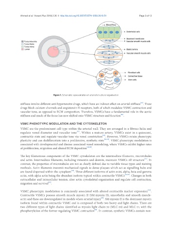
Figure 1. Schematic representation of arterial structural organisation
[25]
stiffness involve different anti-hypertensive drugs, which have an indirect effect on arterial stiffness . These
drugs block calcium-channels and angiotensin II receptors, both of which modulate VSMC contraction and
vascular tone, as opposed to ECM composition. Therefore, VSMCs have a fundamental role in the aortic
[26]
stiffness and much of the focus has now shifted onto VSMC structure and function .
VSMC PHENOTYPIC MODULATION AND THE CYTOSKELETON
VSMC are the predominant cell type within the arterial wall. They are arranged in a fibrous helix and
[27]
regulate vessel diameter and vascular tone . Within a mature artery, VSMCs exist in a quiescent,
[28]
contractile state and regulate vascular tone via vessel constriction . However, VSMCs retain phenotypic
plasticity and can dedifferentiate into a proliferative, synthetic state [29,30] . VSMC phenotypic modulation is
associated with developmental and disease associated vessel remodeling, where VSMCs exhibit higher rates
of proliferation, migration and altered ECM deposition [29,30] .
The key filamentous components of the VSMC cytoskeleton are the intermediate filaments, microtubules
[31]
and actin. Intermediate filaments, including vimentin and desmin, maintain VSMCs 3D structure . In
contrast, the properties of microtubules are not as clearly defined due to variable tissue types and staining
methods. Actin filaments transmit mechanical signals to dense plaques which act as signalling hubs and
[24]
are found dispersed within the cytoplasm . Three different isoforms of actin exist, alpha, beta and gamma
actin, with alpha actin being the abundant isoform typical within contractile VSMCs [24,32] . Changes in both
extracellular and intracellular tension, alter actin cytoskeletal organisation and regulate cell contraction,
[33]
migration and survival .
[34]
VSMC phenotypic modulation is commonly associated with altered contractile marker expression .
Contractile VSMCs possess smooth muscle myosin II (SM-myosin II), smoothelin and smooth muscle-
[35]
actin and these are downregulated in models where arterial injury . SM-myosin II is the dominant myosin
isoform found within contractile VSMC and is composed of both two heavy and light chains. There are
two different types of light chains identified as myosin light chain-20 (MLC-20) and MLC-17, with the
[36]
phosphorylation of the former regulating VSMC contraction . In contrast, synthetic VSMCs contain non-