
Page 75 - Read Online
P. 75
Squizzato et al. Vessel Plus 2023;7:16 https://dx.doi.org/10.20517/2574-1209.2023.05 Page 3 of 14
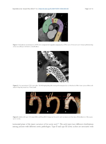
Figure 1. Multiplanar reconstruction (MPR) of a computed tomography angiography (CTA) scan of the aortic arch showing the landing
zones according to Ishimaru’s classification.
Figure 2. Post-procedural CTA scan after TEVAR highlighting the endograft malapposition at the level of the inner curve of the arch
determining the presence of “bird beak”.
Figure 3. Aortic arch type I (A), type II (B), and Type III (C) based on the aortic arch curvature and the take-off distribution of the supra-
aortic trunks.
[13]
horizontal plane of the inner curvature of the aortic arch . The arch types have different distributions
among patients with different aortic pathologies. Type II and type III aortic arches are associated with