
Page 229 - Read Online
P. 229
Somers et al. Vessel Plus 2024;8:15 https://dx.doi.org/10.20517/2574-1209.2023.48 Page 3 of 9
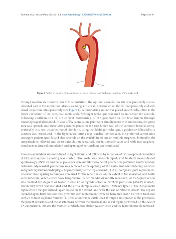
Figure 1. Preferred location of a non-dissected part of the aorta at the lesser curvature of the aortic arch.
through median sternotomy. For DA cannulation, the optimal cannulation site was preferably a non-
dissected part in the anterior or lateral ascending aortic wall, determined on the CT preoperatively and with
visual inspection intraoperatively (see Figure 1). A purse-string suture was placed superficially, often in the
lesser curvature of the proximal aortic arch. Seldinger technique was used to introduce the cannula,
following confirmation of the correct positioning of the guidewire in the true lumen through
transesophageal ultrasound. In case of FA cannulation, prior to or simultaneous with sternotomy, the groin
area was opened, and purse-string sutures placed in the true lumen wall of the common femoral artery,
preferably in a non-dissected vessel. Similarly, using the Seldinger technique, a guidewire followed by a
cannula was introduced. In the hyperacute setting (e.g., cardiac tamponade), the preferred cannulation
strategy is patient-specific and also depends on the availability of one or multiple surgeons. Preferably, the
tamponade is relieved and direct cannulation is started, but in unstable cases and with two surgeons,
simultaneous femoral cannulation and opening of pericardium can be initiated.
Venous cannulation was introduced in right atrium and followed by initiation of extracorporeal circulation
(ECC) and systemic cooling was started. The aorta was cross-clamped, and bilateral near-infrared
spectroscopy (INVOS) and radial pressures were monitored to detect possible malperfusion and/or cerebral
ischemia. Myocardial protection was achieved after opening of the aorta and administering selective
antegrade crystalloid cardioplegia. Supracoronary aortic replacement (SCAR), composite graft replacement,
or aortic valve-sparing techniques were used for the repair based on the extent of the dissection and aortic
valve function. When a core body temperature (either bladder or rectally measured) of 25 degrees or less
was reached (20 degrees or lower in case no antegrade selective cerebral perfusion [ASCP] is used),
circulatory arrest was initiated and the cross-clamp released unless DeBakey type II. The distal aortic
replacement was performed, again based on the extent, and with the use of bilateral ASCP. The repairs
included open distal anastomosis, proximal arch replacement (zone 0), hemiarch (zone 1 or 2) or total arch
with or without elephant trunk (ET). Circulation was re-established through a side branch of the prosthesis,
the patient rewarmed and the anastomosis between the proximal and distal repair performed. In the case of
FA cannulation, this was the moment at which cannulation was switched and the femoral cannula removed.