
Page 115 - Read Online
P. 115
Page 8 of 14 Monaco et al. Vessel Plus 2023;7:23 https://dx.doi.org/10.20517/2574-1209.2023.113
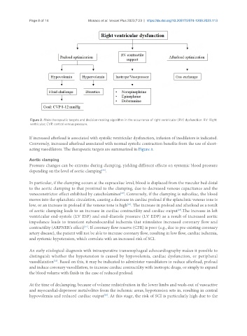
Figure 2. Main therapeutic targets and decision-making algorithm in the occurrence of right ventricular (RV) dysfunction. RV: Right
ventricular; CVP: central venous pressure.
If increased afterload is associated with systolic ventricular dysfunction, infusion of inodilators is indicated.
Conversely, increased afterload associated with normal systolic contraction benefits from the use of short-
acting vasodilators. The therapeutic targets are summarized in Figure 3.
Aortic clamping
Pressure changes can be extreme during clamping, yielding different effects on systemic blood pressure
depending on the level of aortic clamping .
[28]
In particular, if the clamping occurs at the supraceliac level, blood is displaced from the vascular bed distal
to the aortic clamping to that proximal to the clamping, due to decreased venous capacitance and the
[29]
venoconstrictor effect exhibited by catecholamines . Conversely, if the clamping is subceliac, the blood
moves into the splanchnic circulation, causing a decrease in cardiac preload if the splanchnic venous tone is
[30]
low, or an increase in preload if the venous tone is high . The increase in preload and afterload as a result
of aortic clamping leads to an increase in cardiac contractility and cardiac output .The increase in left
[29]
ventricular end-systole (LV ESP) and end-diastole pressure (LV EDP) as a result of increased aortic
impedance leads to transient subendocardial ischemia that stimulates increased coronary flow and
contractility (ARPNER’s effect) . If coronary flow reserve (CFR) is poor (e.g., due to pre-existing coronary
[31]
artery disease), the patient will not be able to increase coronary flow, resulting in low flow, cardiac ischemia,
and systemic hypotension, which correlate with an increased risk of SCI.
An early etiological diagnosis with intraoperative transesophageal echocardiography makes it possible to
distinguish whether the hypotension is caused by hypovolemia, cardiac dysfunction, or peripheral
[32]
vasodilatation . Based on this, it may be indicated to administer vasodilators to reduce afterload, preload
and induce coronary vasodilation, to increase cardiac contractility with inotropic drugs, or simply to expand
the blood volume with fluids in the case of reduced preload.
At the time of declamping, because of volume redistribution in the lower limbs and wash-out of vasoactive
and myocardial-depressor metabolites from the ischemic areas, hypotension sets in, resulting in central
hypovolemia and reduced cardiac output . At this stage, the risk of SCI is particularly high due to the
[28]