
Page 22 - Read Online
P. 22
Page 6 of 10 Korkmaz et al. Rare Dis Orphan Drugs J 2022;1:16 https://dx.doi.org/10.20517/rdodj.2022.26
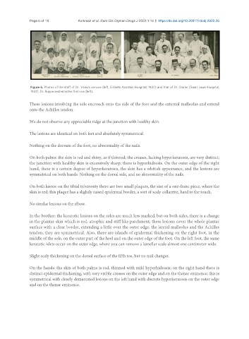
Figure 6. Photos of the staff of Dr. Veau’s service (left, Enfants Assistés Hospital, 1922) and that of Dr. Darier (Saint Louis Hospital,
1922). Dr. Ruppe seated in the first row (left).
These lesions involving the sole encroach onto the side of the foot and the external malleolus and extend
onto the Achilles tendon.
We do not observe any appreciable ridge at the junction with healthy skin.
The lesions are identical on both feet and absolutely symmetrical.
Nothing on the dorsum of the foot, no abnormality of the nails.
On both palms: the skin is red and shiny, as if thinned; the creases, lacking hyperkeratosis, are very distinct;
the junction with healthy skin is excessively sharp; there is hyperhidrosis. On the outer edge of the right
hand, there is a certain degree of hyperkeratosis, the skin has a whitish appearance, and the lesions are
symmetrical on both hands. Nothing on the dorsal side, and no abnormality of the nails.
On both knees: on the tibial tuberosity there are two small plaques, the size of a one-franc piece, where the
skin is red; this plaque has a slightly raised epidermal border, a sort of scaly collarette, hard to the touch.
No similar lesions on the elbow.
In the brother: the keratotic lesions on the soles are much less marked, but on both sides, there is a change
in the plantar skin which is red, atrophic and stiff like parchment; these lesions cover the whole plantar
surface with a clear border, extending a little over the outer edge, the lateral malleolus and the Achilles
tendon; they are symmetrical. Also, there are islands of epidermal thickening on the right foot, in the
middle of the sole, on the outer part of the heel and on the outer edge of the foot. On the left foot, the same
keratotic islets occur on the outer edge, where you can remove a lamellar scale almost one centimeter wide.
Slight scaly thickening on the dorsal surface of the fifth toe, but no nail changes.
On the hands: the skin of both palms is red, thinned with mild hyperhidrosis; on the right hand there is
distinct epidermal thickening, with very visible creases on the outer edge and on the thenar eminence; this is
symmetrical with clearly demarcated lesions on the left hand with discrete hyperkeratosis on the outer edge
and on the thenar eminence.