
Page 758 - Read Online
P. 758
Page 8 of 13 Murthy et al. Plast Aesthet Res 2020;7:64 I http://dx.doi.org/10.20517/2347-9264.2020.72
A B C
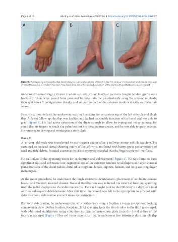
Figure 5. Appearance of reconstructed hand following suction lipectomy of the ALT flap for contour improvement and regular sessions
of hand therapy (A-C). Patient has pain-free, functional arc of flexion and extension of the digits with aesthetically pleasing result
underwent second stage extensor tendon reconstruction. Bilateral palmaris longus tendon grafts were
harvested. These were passed from proximal to distal into the pseudosheath using the silicone implants,
then split into a Y configuration distally, and sutured to each of the extensor tendons distally via Pulvertaft
weave.
Finally, six months later, he underwent suction lipectomy for re-contouring of the left anterolateral thigh
flap. At latest follow up, the flap was healthy, and he had reasonable function of the hand and was able to
grip [Figure 5]. He had active extension of the digits enough to allow for typing and video-gaming. He
could flex his fingers to touch the palm but not the distal palmar crease, and he was able to grasp objects.
He returned to driving and working as a store clerk.
Case 2
A 47-year-old male was transferred to our trauma center after a rollover motor vehicle accident. He
sustained an isolated dorsal shearing injury of the left wrist and hand with heavy, gross contamination of
road and field debris. Focused examination of the extremity revealed that the fingers were well-perfused.
He was taken to the operating room for exploration and debridement [Figure 6]. He was found to have
significant skin and soft tissue loss; segmental loss of the extensor tendons to all fingers; and open coronal
plane fractures of the distal radius, distal ulna, scaphoid, lunate, capitate, hamate, and long and ring finger
metacarpals.
At the index procedure, he underwent thorough excisional debridement, placement of antibiotic cement
beads, and vacuum-assisted closure. Skeletal stabilization was achieved via external fixation, spanning
from the radial diaphysis to the index metacarpal. He was brought back to the OR every 2-3 days for a total
of three subsequent debridements. After this time, the wound was felt to be appropriate to proceed with
definitive bony stabilization and soft tissue reconstruction.
For bony stabilization, he underwent total wrist arthrodesis using a Synthes 3.5-mm metaphyseal locking
compression plate (DePuy Synthes, Raynham, MA) spanning from the distal radius to the third metacarpal,
with additional stabilization using a Synthes 2.7-mm reconstruction plate from the distal radius to the
fourth metacarpal [Figure 7] For soft tissue reconstruction, he underwent free latissimus dorsi muscle flap