
Page 702 - Read Online
P. 702
Page 2 of 12 Laplant et al. Plast Aesthet Res 2020;7:60 I http://dx.doi.org/10.20517/2347-9264.2020.69
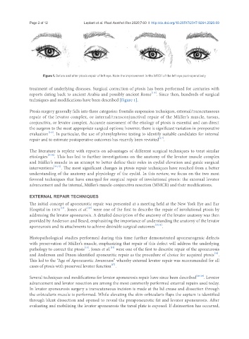
Figure 1. Before and after ptosis repair of left eye. Note the improvement in the MRD1 of the left eye post-operatively
treatment of underlying diseases. Surgical correction of ptosis has been performed for centuries with
[1,2]
reports dating back to ancient Arabia and possibly ancient Rome . Since then, hundreds of surgical
techniques and modifications have been described [Figure 1].
Ptosis surgery generally falls into three categories: frontalis suspension techniques, external/transcutaneous
repair of the levator complex, or internal/transconjunctival repair of the Müller’s muscle, tarsus,
conjunctiva, or levator complex. Accurate assessment of the etiology of ptosis is essential and can direct
the surgeon to the most appropriate surgical options; however, there is significant variation in preoperative
[3-5]
evaluation . In particular, the use of phenylephrine testing to identify suitable candidates for internal
[6,7]
repair and to estimate postoperative outcomes has recently been revisited .
The literature is replete with reports on advantages of different surgical techniques to treat similar
etiologies [8-10] . This has led to further investigations on the anatomy of the levator muscle complex
and Müller’s muscle in an attempt to better define their roles in eyelid elevation and guide surgical
interventions [11,12] . The most significant changes in ptosis repair techniques have resulted from a better
understanding of the anatomy and physiology of the eyelid. In this review, we focus on the two most
favored techniques that have emerged for surgical repair of involutional ptosis: the external levator
advancement and the internal, Müller’s muscle-conjunctiva resection (MMCR) and their modifications.
EXTERNAL REPAIR TECHNIQUES
The initial concept of aponeurotic repair was presented at a meeting held at the New York Eye and Ear
[13]
[13]
Hospital in 1970 . Jones et al. were one of the first to describe the repair of involutional ptosis by
addressing the levator aponeurosis. A detailed description of the anatomy of the levator anatomy was then
provided by Anderson and Beard, emphasizing the importance of understanding the anatomy of the levator
aponeurosis and its attachments to achieve desirable surgical outcomes [13,14] .
Histopathological studies performed during this time further demonstrated aponeurogenic defects
with preservation of Müller’s muscle, emphasizing that repair of this defect will address the underlying
[15]
[13]
pathology to correct the ptosis . Jones et al. were one of the first to describe repair of the aponeurosis
[16]
and Anderson and Dixon identified aponeuritic repair as the procedure of choice for acquired ptosis .
This led to the “Age of Aponeurotic Awareness” whereby external levator repair was recommended for all
[17]
cases of ptosis with preserved levator function .
Several techniques and modifications for levator aponeurosis repair have since been described [18-20] . Levator
advancement and levator resection are among the most commonly performed external repairs used today.
In levator aponeurosis surgery a transcutaneous incision is made at the lid crease and dissection through
the orbicularis muscle is performed. While elevating the skin-orbicularis flaps the septum is identified
through blunt dissection and opened to reveal the preaponeurotic fat and levator aponeurosis. After
evaluating and mobilizing the levator aponeurosis the tarsal plate is exposed. If disinsertion has occurred,