Page 324 - Read Online
P. 324
Lasso. Plast Aesthet Res 2020;7:30  I  http://dx.doi.org/10.20517/2347-9264.2019.75                                                   Page 7 of 12

               Table 3. Fluorescence patterns are registered according to the images precepted 15 min after ICG injection
                     PRE-LVA fluorescence pattern   POST-LVA fluorescence   Baseline SPECT-CT/  Post LVA SPECT-CT/
                                                  pattern          lymphoscintigraphy   lymphoscintigraphy
                1        Diffuse/stardust  Stardust                      -                    -
                2        Diffuse/stardust   Diffuse/linear in some areas   -                  ++
                3        Diffuse/stardust  Stardust                      -                    +
                4        Stardust          Linear                        -                     +*
                5        Stardust          Linear                        -                    +
                6        Linear, stardust  Linear - slow progression     -                    +
                7        Linear/stardust   Linear                        -                    ++
                8        Diffuse           Diffuse                       -                    +
                9        Stardust/splash   Splash                        -                    +
                10       Linear/splash     Linear                        +                    +
                11       Linear/stardust   Linear                        -                    ++
                12       Diffuse           Diffuse                       -                    +
                13       Splash            Splash                        -                    -
                14       Stardust          Linear/splash                 -                    ++*
                15       Diffuse/splash    Splash                        -                    +
                16       Diffuse           Linear/diffuse                -                    -
                17       Linear/stardust   Linear                        -                    ++
                18       Stardust          Stardust                      -                    +
                19       Linear/splash     Linear                        -                    +
                20       Splash            Splash                        -                    +
               SPECT-CT/lymphoscintigraphy results are described at baseline and 1 year after lymphovenous anastomosis (LVA). Results are marked
               as follows: -: No detectable migration of the tracer from the site of injection to the groin or axilla, lymphatic leakage or dermal backflow; +:
               The report of new images of lymphatic flow along the limb; ++: The presence of lymph nodes at any point in the limb; *The presence of
               extraanatomical lymphatic flow; Note that fluorescence patterns are detected 15 min after injection and SPECT-CT/lymphoscintigraphy
               images are obtained 3 h after injection. This evaluation was performed the day before LVA and 1 year after it. Patterns are labeled
               according to Yamamoto’s dermal backflow classification [11]


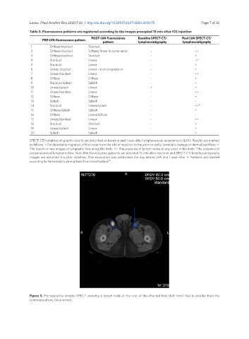
               Figure 3. Pre-operative lympho-SPECT showing a lymph node at the root of the affected limb (left limb) that is smaller than the
               contralateral one (blue arrow).

