
Page 269 - Read Online
P. 269
Page 6 of 13 Cohen-Shohet et al. Plast Aesthet Res 2019;5:28 I http://dx.doi.org/10.20517/2347-9264.2019.030
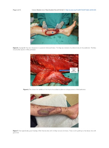
Figure 5. A propeller flap was designed on a posterior tibial perforator. The flap was isolated circumferentially on the perforator. The blue
X marks the location of the perforator
Figure 6. This shows the isolation of the flap in the subfascial plane on a large posterior tibial perforator
Figure 7. Post-operatively, good healing of the fracture site with no flap necrosis is shown. There is skin grafting to the donor site with
good take