
Page 261 - Read Online
P. 261
Page 8 of 10 Scaglioni et al. Plast Aesthet Res 2019;6:27 I http://dx.doi.org/10.20517/2347-9264.2019.41
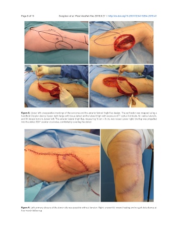
Figure 8. Upper left: preoperative markings of the sarcoma and the anterior lateral thigh flap design. The perforator was mapped using a
handheld Doppler device. Upper right: large soft tissue defect on the lateral thigh with exposure of T ractus iliotibialis, M. vastus lateralis,
and M. biceps femoris. Lower left: The anterior lateral thigh flap, measuring 16 cm × 8 cm, was raised. Lower right: the flap was propelled
into the defect 130° counter clockwise, comfortably covering the defect
Figure 9. Left: primary closure of the donor site was possible without tension. Right: uneventful wound healing and no gait disturbance at
five-month follow-up