
Page 249 - Read Online
P. 249
O’Connor et al. Plast Aesthet Res 2019;6:26 I http://dx.doi.org/10.20517/2347-9264.2019.38 Page 5 of 9
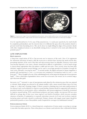
A B
Figure 1. A: laparoscopic image of bowel herniated above the posterior rectus sheath and in contact with the exposed mesh. B: CT image
showing the dehisced posterior rectus sheath with bowel herniated above. Linea alba is intact with no full thickness herniation
time for reperitonealization. In the case of late presentations, 2-3 weeks post operatively, the mesh should
be completely peritonealized, and the objective here is to obliterate the space above the posterior sheath to
prevent bowel from herniating above. This can be done by simply lifting the edges of the posterior sheath
with either transfascial sutures or an absorbable tacking device to attach it to the mesh and obliterate the
[27]
potential space .
LATE COMPLICATIONS
Mesh exposure
A long-term complication of SSI or flap necrosis can be exposure of the mesh. One of the arguments
for retrorectus placement of mesh is that the rectus acts as another layer between the mesh and the skin,
preventing exposure of the mesh if the skin and subcutaneous tissue are debrided. However, when mesh
does become exposed, it can create a chronic wound that is difficult to epithelialize. A trial of mesh salvage
is almost always appropriate when the patient is stable and not septic. Once necrotic tissue has been fully
debrided, negative pressure wound therapy can be used to encourage granulation formation through the
[28]
mesh pores, ultimately covering the mesh . In animal models, the degree and speed of tissue integration is
greatest with macroporous (1.8 mm × 3.4 mm pore size) compared to microporous mesh (0.9 mm × 1 mm
[29]
pore size) . This is thought to be one of the contributing factors in the improved salvage rate of macroporous
[12]
mesh . Once a thick base of granulation tissue covers the entire mesh, the wound can be covered using a
split-thickness skin graft.
[13]
Berrevoet et al. reviewed 54 cases of macroporous mesh placed in the retrorectus space that was exposed
or infected. Mesh salvage was achieved in all patients studied, with a mean wound closure time of 44 days
(range, 26-73 days) and 5 dressing changes. If chronic wounds or draining sinus tracts exist, small portions of
the exposed mesh can be debrided to improve wound healing. Patients should be supported with enteral or
parenteral nutrition to avoid protein calorie malnutrition, and immunosuppressants should be minimized
to improve wound healing. PTFE, multifilament polyester, and heavy weight, microporous PPE are more
difficult to salvage due to their poor tissue ingrowth and biofilm formation, which prevents complete
clearance of bacteria. If explantation is required, meticulous removal of all mesh and synthetic material,
including suture and tacks, should be performed. Primary closure with staged repeat repair should be
[9]
performed if possible, or use of biosynthetic mesh if required for coverage of intrabdominal contents .
Enterocutaneous fistula
Enterocutaneous fistula (ECF) is a feared long-term complication of hernia repair, occurring on average
2 years after the index operation. These often present as a chronic mesh infection that is followed by bilious