
Page 152 - Read Online
P. 152
Göksel et al. Plast Aesthet Res 2019;6:17 I http://dx.doi.org/10.20517/2347-9264.2019.12 Page 5 of 8
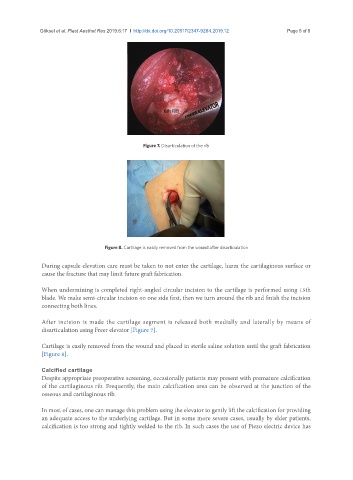
Figure 7. Disarticulation of the rib
Figure 8. Cartilage is easily removed from the wound after disarticulation
During capsule elevation care must be taken to not enter the cartilage, harm the cartilaginous surface or
cause the fracture that may limit future graft fabrication.
When undermining is completed right-angled circular incision to the cartilage is performed using 15th
blade. We make semi-circular incision on one side first, then we turn around the rib and finish the incision
connecting both lines.
After incision is made the cartilage segment is released both medially and laterally by means of
disarticulation using Freer elevator [Figure 7].
Cartilage is easily removed from the wound and placed in sterile saline solution until the graft fabrication
[Figure 8].
Calcified cartilage
Despite appropriate preoperative screening, occasionally patients may present with premature calcification
of the cartilaginous rib. Frequently, the main calcification area can be observed at the junction of the
osseous and cartilaginous rib.
In most of cases, one can manage this problem using the elevator to gently lift the calcification for providing
an adequate access to the underlying cartilage. But in some more severe cases, usually by elder patients,
calcification is too strong and tightly welded to the rib. In such cases the use of Piezo electric device has