
Page 76 - Read Online
P. 76
Bonomi et al. Plast Aesthet Res 2018;5:8 I http://dx.doi.org/10.20517/2347-9264.2017.93 Page 3 of 8
Figure 1. A 48-year-old man with a dermatofibrosarcoma protuberans measuring 16 cm × 14 cm × 6 cm was present on the mid back
A B
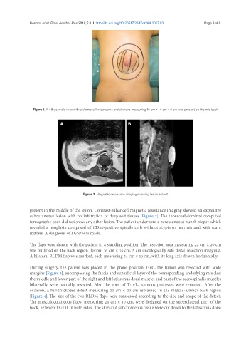
Figure 2. Magnetic resonance imaging showing lesion extent
present in the middle of the lesion. Contrast-enhanced magnetic resonance imaging showed an expansive
subcutaneous lesion with no infiltration of deep soft tissues [Figure 2]. The thoracoabdominal computed
tomography-scan did not show any other lesion. The patient underwent a percutaneous punch biopsy which
revealed a neoplasia composed of CD34-positive spindle cells without atypia or necrosis and with scant
mitosis. A diagnosis of DFSP was made.
The flaps were drawn with the patient in a standing position. The resection area measuring 22 cm × 20 cm
was outlined on the back region (lesion: 16 cm × 14 cm, 3 cm oncologically safe distal resection margins).
A bilateral RLDM flap was marked, each measuring 24 cm × 10 cm, with its long axis drawn horizontally.
During surgery, the patient was placed in the prone position. First, the tumor was resected with wide
margins [Figure 3], encompassing the fascia and superficial layer of the corresponding underlying muscles:
the middle and lower part of the right and left latissimus dorsi muscle, and part of the sacrospinalis muscles
bilaterally were partially resected. Also the apex of T10-L2 spinous processes were removed. After the
excision, a full-thickness defect measuring 22 cm × 20 cm remained in the middle-lumbar back region
[Figure 4]. The size of the two RLDM flaps were reassessed according to the size and shape of the defect.
The musculocutaneous flaps, measuring 24 cm × 10 cm, were designed on the superolateral part of the
back, between T8-T11 in both sides. The skin and subcutaneous tissue were cut down to the latissimus dorsi