
Page 38 - Read Online
P. 38
Kannan et al. Plast Aesthet Res 2018;5:4 I http://dx.doi.org/10.20517/2347-9264.2018.02 Page 3 of 6
Figure 1. A graphical illustration of the labio-mandibular flap raised from the peri-oral and lower lip areas, along the LMF and vermilion
border, illustrated by “1”. Note also the Y-V advancement of the oral commissure into the flap itself (illustrated as “2”). The presence of
the orbicularis oris musculocutaneous perforator within the distal flap ensures its continued perfusion while the narrow cutaneous bridge
at the oral commissure suffices for venous and lymphatic return
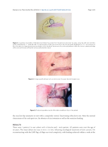
Figure 2. A large superficial basal cell carcinoma over the upper lip/commissural area
Figure 3. Final post-operative results of the labio-mandibular flap on the patient
the excellent lip symmetry at rest with a completely normal functioning orbicularis oris. Note the normal
dimensions of the oral aperture, the absence of microstomia as well as the seamless healing.
RESULTS
There were 7 patients’ in our cohort with 6 females and 1 male patient. All patients were over the age of
60 years. The mean defect size was 23 mm × 22 mm, following oncological resections of skin cancers. On
reconstructing with the LMF flap, all flaps survived completely, with healing achieved within a week at the