
Page 333 - Read Online
P. 333
Page 4 of 14 Khan. Plast Aesthet Res 2018;5:45 I http://dx.doi.org/10.20517/2347-9264.2018.58
A B C
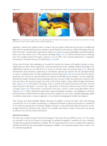
Figure 2. Picture showing showing implant in muscle split pocket (A); illustrations showing level of muscle split at the junction of middle
and lower third sternum and implant in muscle splitting pocket (B, C)
quadrant, a muscle split biplane pocket is created. The pectoralis is split from the junction of middle and
lower third of sternal attachment and continues up and laterally to the anterior axillary fold along a line just
below Neo-NAC . Round cohesive gel silicone implants are placed in muscle split biplane pocket through the
lower end of the vertical scar or Wise pattern markings [Figure 2A-C]. Patients with periareolar markings
have their implants placed through a separate access in IMC. Once implant placement is completed,
haemostasis is checked and access is closed [Figure 3A and B].
Using Lane’s forceps, skin markings are checked for tension free closure with implant already in place.
Adjustments are made where required for a safe and tension free closure. Marked medially based flap is de-
epithelialized, leaving 4.2 cm wide NAC as a routine in all three types of markings [Figure 4A]. Skin and
subcutaneous tissue excision is performed, in moderation, superior and lateral to the de-epithelialised flap
to create an adequate space for flap mobilisation and resetting [Figure 4B]. In vertical and Wise pattern
markings, skin excision is continued below the medially based flap and according to the skin markings.
Between the medial and lateral vertical markings, the tissue excision is little more generous to prevent lower
pole redundancy but leaving enough tissue layer and without implant being visible through intermediate
layer [Figure 5A]. In Wise pattern markings, tissue is excised from lateral and medial extensions, into the
respective pole of the breast, in a similar way. A good layer of breast parenchymal tissue is left for implant
coverage [Figure 5B]. Haemostasis is performed, three layer closure is done using absorbable sutures
[Figures 6 and 7]. Flap is checked for tension free closure and nipple circulation, if any tightness is observed,
piecemeal tissue is removed between flap and new-NAC margins. In case of skin envelope tightening, nipple
circulation compromise or venous congestion, I do not hesitate exchanging for smaller size implants.
After closure, steri-strips and light adhesive dressings are applied. A decent size hole is left in the dressing
covering NAC for its circulation monitoring. A compression brassiere is placed and patients are transferred
to the ward for postoperative monitoring. Nipple circulation is checked hourly before the patients are dis-
charged. Patients are advised to take a picture of the nipples and send it to author if they notice any change
in colour for an early and timely intervention.
Statistical analysis
The data were analysed using the Statistical Package for the Social Sciences (SPSS), version 19.0. The results
are presented in the text as frequency, percentage for qualitative/categorical variables and mean, Standard
deviation for quantitative/continuous variables. The Chi-square/exact test is used to compare the categorical
variables and ANOVA test for quantitative/continuous variables. In all statistical analysis, only P-values < 0.05
are considered significant.