
Page 297 - Read Online
P. 297
Madiedo et al. Plast Aesthet Res 2018;5:40 I http://dx.doi.org/10.20517/2347-9264.2018.40 Page 5 of 10
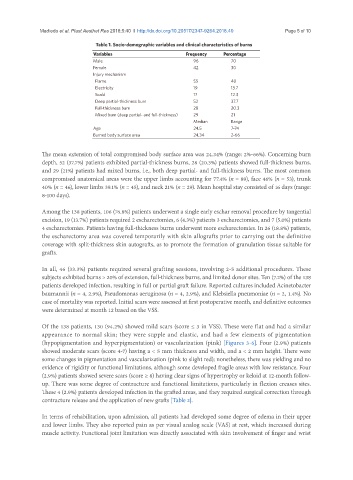
Table 1. Socio-demographic variables and clinical characteristics of burns
Variables Frequency Percentage
Male 96 70
Female 42 30
Injury mechanism
Flame 55 40
Electricity 19 13.7
Scald 17 12.3
Deep partial-thickness burn 52 37.7
Full-thickness burn 28 20.3
Mixed burn (deep partial- and full-thickness) 29 21
Median Range
Age 24.5 7-74
Burned body surface area 24.34 2-66
The mean extension of total compromised body surface area was 24.34% (range: 2%-66%). Concerning burn
depth, 52 (37.7%) patients exhibited partial-thickness burns, 28 (20.3%) patients showed full-thickness burns,
and 29 (21%) patients had mixed burns, i.e., both deep partial- and full-thickness burns. The most common
compromised anatomical areas were the upper limbs accounting for 77.4% (n = 89), face 46% (n = 53), trunk
40% (n = 46), lower limbs 39.1% (n = 45), and neck 21% (n = 29). Mean hospital stay consisted of 16 days (range:
8-100 days).
Among the 138 patients, 106 (76.8%) patients underwent a single early eschar removal procedure by tangential
excision, 19 (13.7%) patients required 2 escharectomies, 6 (4.3%) patients 3 escharectomies, and 7 (5.0%) patients
4 escharectomies. Patients having full-thickness burns underwent more escharectomies. In 26 (18.8%) patients,
the escharectomy area was covered temporarily with skin allografts prior to carrying out the definitive
coverage with split-thickness skin autografts, as to promote the formation of granulation tissue suitable for
grafts.
In all, 46 (33.3%) patients required several grafting sessions, involving 2-5 additional procedures. These
subjects exhibited burns > 20% of extension, full-thickness burns, and limited donor sites. Ten (7.2%) of the 138
patients developed infection, resulting in full or partial graft failure. Reported cultures included Acinetobacter
baumannii (n = 4, 2.9%), Pseudomonas aeruginosa (n = 4, 2.9%), and Klebsiella pneumoniae (n = 2, 1.4%). No
case of mortality was reported. Initial scars were assessed at first postoperative month, and definitive outcomes
were determined at month 12 based on the VSS.
Of the 138 patients, 130 (94.2%) showed mild scars (score ≤ 3 in VSS). These were flat and had a similar
appearance to normal skin; they were supple and elastic, and had a few elements of pigmentation
(hypopigmentation and hyperpigmentation) or vascularization (pink) [Figures 3-6]. Four (2.9%) patients
showed moderate scars (score 4-7) having a < 5 mm thickness and width, and a < 2 mm height. There were
some changes in pigmentation and vascularization (pink to slight red); nonetheless, there was yielding and no
evidence of rigidity or functional limitations, although some developed fragile areas with low resistance. Four
(2.9%) patients showed severe scars (score ≥ 8) having clear signs of hypertrophy or keloid at 12-month follow-
up. There was some degree of contracture and functional limitations, particularly in flexion creases sites.
These 4 (2.9%) patients developed infection in the grafted areas, and they required surgical correction through
contracture release and the application of new grafts [Table 2].
In terms of rehabilitation, upon admission, all patients had developed some degree of edema in their upper
and lower limbs. They also reported pain as per visual analog scale (VAS) at rest, which increased during
muscle activity. Functional joint limitation was directly associated with skin involvement of finger and wrist