
Page 28 - Read Online
P. 28
Azadgoli et al. Plast Aesthet Res 2018;5:3 I http://dx.doi.org/10.20517/2347-9264.2017.32 Page 5 of 12
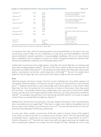
Zaretski et al. [33] 2004 Femur Free vascularized fibula flap ± allograft
Tibia Free double-barreled fibula
Capanna et al. [34] 1993 Femur Free vascularized fibula flap + allograft
Tibia
Beris et al. [35] 2011 Femur Free vascularized fibula ± allograft
Tibia Free double-barreled fibula
Yajima and Tamai [36] 1994 Femur Twin-barrelled vascularized fibular graft
Tibia
Ankle
Duffy et al. [37] 2000 Femur Free vascularized fibula
Tibia
Rush and Koman [38] 1997 Tibia Fibula-flexor halluces longus osteomuscular
flap
Mastorakos et al. [39] 2002 Tibia LD
RA
Gastrocnemius-Soleus
Gastrocnemius-RA
Doi et al. [41] 1998 Lower leg Gracilis + motor nerve
Doi et al. [42] 1999 Thigh LD + motor nerve
Lower leg Gracilis + motor nerve
LD: latissimus dorsi; RA: rectus abdominis; ALT: anterior lateral thigh
free latissimus dorsi flaps, while the remaining patients received pedicled flaps. In the patients who were
reconstructed using free flaps, the only complications were leg edema and mild lymphedema, which the
authors attributed to ischemic reperfusion or venous/lymphatic insufficiency. The use of a myocutaneous
flap in combination with an autologous vein graft also results in decreased postoperative infection rates,
[14]
treatment of lymphedema and fistula, and increased graft patency rates .
Aesthetically, reconstruction of the thigh requires a large flap with muscle bulk that can eliminate dead
[10]
space while providing adequate contour . The use of a free rectus abdominis flap has been reported to be
particularly successful for this purpose [6,10] . The latissimus dorsi flap, which is thin, large with a long vascular
pedicle, ± neurotization has also been used for large defects of the thigh [6,10] . The use of the anterior lateral
[15]
thigh (ALT) flap for large thigh defects, particularly of the posterior thigh, has also been reported .
Knee
Obtaining adequate soft tissue coverage of the knee remains challenging for many plastic surgeons, not
only because of the biomechanics of the knee, but also due to exposure of vital structures as well as the joint
space [16-18] . Rotational muscle flaps or myocutaneous flaps such as gastrocnemius or reverse anterior lateral
thigh flaps have been the mainstay for the reconstruction of tumors in this location. These flaps usually
have low donor - site morbidity. However more complex defects may require the use of free tissue transfer.
In these cases the deep-seated recipient popliteal vessels of the knee can make microvascular anastomosis
[19]
difficult , an autologous vein graft loop can be used and the distal SFA and SFV can be used as recipient
[14]
vessels if there is an extended field of neoadjuvant radiation .
Multiple donor sites have been successful used in free flap coverage of knee defects. These include latissimus
[6]
dorsi, rectus abdominis, and scapula flaps . When there is a large contour defect in the popliteal fossa that
[10]
does not require much filling of the muscular space, Leow et al. have also described the use of a free mini-
transverse rectus abdominis (TRAM) myocutaneous flap.
In many cases where complex reconstruction of the knee region is needed, salvage of the popliteal
artery, which can often be involved in the disease process, becomes critical. This has traditionally been
[20]
accomplished using a combination of a local gastrocnemius flap with an interpositional vein graft .
[21]
However, Miyamoto et al. described two cases of successful one-stage reconstruction of complex knee
defects including the popliteal artery using a free flow-through ALT flap. Although the use of a deep