
Page 206 - Read Online
P. 206
Garlick et al. Plast Aesthet Res 2018;5:29 I http://dx.doi.org/10.20517/2347-9264.2018.36 Page 3 of 5
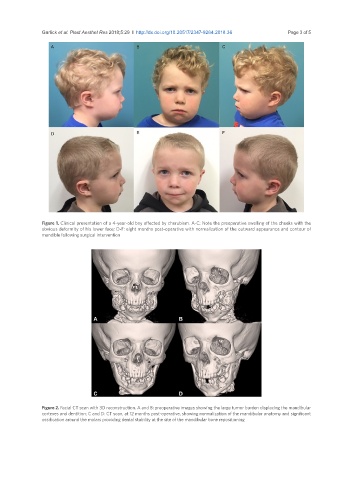
Figure 1. Clinical presentation of a 4-year-old boy affected by cherubism. A-C: Note the preoperative swelling of the cheeks with the
obvious deformity of his lower face; D-F: eight months post-operative with normalization of the outward appearance and contour of
mandible following surgical intervention
Figure 2. Facial CT scan with 3D reconstruction. A and B: preoperative images showing the large tumor burden displacing the mandibular
cortexes and dentition; C and D: CT scan, at 12 months post-operative, showing normalization of the mandibular anatomy and significant
ossification around the molars providing dental stability at the site of the mandibular bone repositioning