
Page 184 - Read Online
P. 184
Page 2 of 4 Flores et al. Plast Aesthet Res 2018;5:24 I http://dx.doi.org/10.20517/2347-9264.2018.31
A B
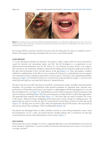
Figure 1. (A) Stevens Johnson syndrome manifested by generalized swelling and full thickness epidermal necrosis of the skin of face
and the mucosa of the lips; (B) oral commissures fusion (arrows) resulting in impairment of normal speech, oral intake, as well as a
compromised aesthetic appearance
the setting of SJS has only been reported four times with this being the first report of complete fusion
[3-6]
(fusion of the upper to lower lip extending all the way to the corner of the mouth).
CASE REPORT
A 19-year-old Hispanic female was referred to the pediatric plastic surgery clinic for severe microstomia
caused by bilateral oral commissure fusion post SJS. The SJS developed as a complication to oral
sulfamethoxazole/trimethoprim used by the patient for the treatment of severe chronic acne vulgaris.
This reaction to the medication resulted in generalized swelling and full thickness epidermal necrosis of
the skin with involvement of face and the mucosa of the lips [Figure 1A]. Once the patient’s condition
stabilized re-epithelization of the affected areas commenced. During the re-epithelization process patient’s
oral commissures fused resulting in impairment of normal speech, oral intake, and compromised aesthetic
appearance [Figure 1B]. Despite the fusion, the involved tissue remained relatively supple, in contrast to the
usual thick and rigid scar associated with thermal or chemical burns).
We report here the successful functional and aesthetic reconstruction using a modified commissuroplasty
technique. The procedure was performed under general anesthesia on outpatient basis. Incisions were
performed at the level of the commissures in the shape of a 4-sided polygons with the long diagonal of 1.5 cm and
the short diagonal of 1 cm [Figure 2A]. Full thickness excision of the scar present was performed preserving
only the deep mucosal lining [Figure 2B]. The exposed mucosa was then divided in a Mercedes-sign pattern.
This generated three separate mucosal flaps [Figure 2C]. The lateral flap was used to resurface the corner
of the mouth, while preventing overlapping sutures lines and re-fusion during the healing process. The
superior flap was used to resurface the upper lip wound and the inferior flap to resurface the lower lip wound
[Figure 2D]. The flaps were secured in place with interrupted deep dermal PDS sutures. The superficial lip
mucosa was closed with a running 6-0 fast absorbing suture [Figure 2E].
The patient was discharged home in stable condition and had an uneventful postoperative course. Upon
the postoperative visit at 6 weeks, the patient showed complete healing with a normalized oral opening,
excellent function and an esthetically pleasant mouth contour [Figure 3].
DISCUSSION
Microstomia has various etiologies. It can be a congenital deformity or an oral manifestation of connective
tissue disorders. Furthermore, it can be secondary to electrical, thermal and caustic injury, or the result of
oncologic resection .
[7]