
Page 209 - Read Online
P. 209
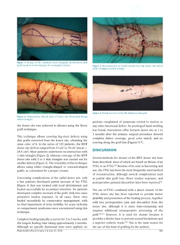
Figure 1: Design of the combined local triangular full-thickness skin
            graft based on 4 skin triangles for rectangular defects  Figure 3: Reconstruction of radial forearm free flap donor site defect
                                                              with 3 triangles (vertical design)


                                                              Figure 4: Partial necrosis of the full-thickness skin graft
            Figure 2: Postoperative clinical views of donor site. Horizontal design
            with 4 triangles
                                                              patients complained of symptoms related to motion or
            the donor site was achieved in allcases using the Iberic   any other functional deficit. No prolonged hand swelling
            graft technique.                                  was found. Assessment ofthe forearm donor site at 1 to
                                                              3 months after the primary surgical procedure showed
            This technique  allows covering big-sized  defects using   complete defect coverage, good color match, and no
            skin grafts extracted from the donor site, obtaining the   scarring along the graft line [Figures 5-7].
            same color of it. In the series of 125 patients, the RFFF
            donor site defects ranged from 15 cm  to 70 cm  (mean ±
                                           2
                                                   2
            24.5 cm ). Most patients underwent reconstruction with   DISCUSSION
                   2
            4 skin triangles [Figure 2], whereas coverage of the RFFF
            donor site with 2 or 3 skin triangles was carried out for   Several methods for closure of the RFFF donor site have
            smaller defects [Figure 3]. The versatility of this technique   been described, most of which are based on theuse of an
                                                                            [12]
            allows  using either triangle-shaped or crescent-shaped   STSG or an FTSG.  Because of its ease in harvesting and
            grafts, as convenient for a proper closure.       use, the STSG has been the most frequently used method
                                                              of reconstruction, although several complications such
            Concerning complications of the radial donor site, only   as  partial skin  graft  loss,  flexor  tendon  exposure,  and
            a  few  patients  developed partial necrosis  of  the  FTSG   postoperative painand discomfort have been reported.
                                                                                                           [9]
            [Figure 4] that was treated with local debridement and
            healed successfully by secondary intention. No patients   The use of FTSG combined with a direct closure of the
            developed complete necrosis of the graft. Only two cases   FTSG donor site  has been  reported to provide better
            presented tendon exposure. In all cases, these sites   pliability and promotion of the healing process, together
            healed secondarily by conservative management,  with   with less postoperative pain and discomfort from the
            no final impairment of wrist mobility. No acute ischemia
            or compartment syndromes were encountered using this   donor  site, although it is more time-consuming  and
            technique.                                        requires additional  intraoperative processing of the
                                                              graft. [28-31]  However, it is used for closure because it
            Complete healing typically occurred for 2 to 3 weeks, with   provides a thicker base to prevent wound breakdown and
                                                                                    [32]
            the longest healing time taking approximately 2 months.   a superior esthetic result.  This is the main reason for
            Although no specific functional tests were applied,  no   the use of this kind of grafting by the authors.
            Plast Aesthet Res || Volume 3 || June 24, 2016                                                199