Page 187 - Read Online
P. 187
decompressions,  medial epicondylectomy,  subcutaneous   these conditions may mimic ulnar nerve syndrome at the
                                              [8]
                        [7]
          transposition, [9]  intramuscular  transposition, [10]  and  wrist, which, in addition to the different symptoms noted
                                [11]
          submuscular transposition.  However, as reported in the   by patients, may render recognition of ulnar neuropathy
          first  description of this neuropathy by Panas  in 1878,   at the elbow difficult.
                                                 [12]
          surgical treatment frequently yields poor results and may
          worsen the initial clinical condition. In a recent study   CAUSES OF FAILURE AFTER PRIMARY
          conducted by the American Association of Hand Surgery,    SURGERY
                                                         [13]
          61% of surgeons reported inferior results following surgical
          ulnar nerve decompression at the elbow compared with   Failure after surgery is mainly due to procedural errors or
          the  results  obtained following decompression  after   technical omissions, frequently represented by incomplete
          carpal tunnel  release  (44% satisfactory  results  vs.  88%,   nerve decompression, failure to recognize nerve instability
          respectively).                                      after the nerve has been decompressed, loosening of
          According to the literature, no surgical technique may be   the nerve anchor after superficial nerve transposition
          defined as superior to the others. [14‑16]  The persistence   with  consequent  spontaneous  nerve  relocation in  the
          of preoperative symptoms after surgery is defined as   epitrochlear‑olecranon channel, perineural fibrosis and
          failure, whereas the reappearance of symptoms after   neurodesis that creates  a new site  of nerve compression
          a  period  of  relief  is  defined  as  recurrence,  and  surgical   in  areas  different  from  those  affected by  the  original
          revision  is  indicated  in  both  cases.   Preoperative   compression,  unintended injury to one or more sensory
                                             [15]
          factors that may be associated with poor results after   regional nerves or to the ulnar nerve itself, articular elbow
          surgery depend on multiple elements, including an   instability due to unintended injury to the ulnar collateral
          incorrect  diagnosis,   advanced  neuropathy  with  a   ligament, or elbow stiffening in the flexed position.
                            [17]
          neurological lesion and muscular atrophy,  coexisting   It is widely accepted  that when primary nerve
                                               [18]
          pathologies such as double crush syndrome,  cervical   decompression is performed, only one of the five possible
                                                 [16]
          spine radiculopathies,  thoracic outlet syndrome,    sites of compression is generally found  to actually  be
                             [19]
                                                         [20]
          ulnar compression syndromes at the wrist, endocrine   responsible  for the  nerve impingement,  and these  sites
                                                                                                [2]
          disorders such as diabetes mellitus or thyroid disease,   are  usually the  epitrochlear‑olecranon channel or the
          and  polyneuropathies,  particularly  if  they  are  associated   Osborne fibrous arcade. The surgeon’s experience will
                                                 [13]
          with muscular atrophy or decreased sensation.  Some of
                                                              generally determine the decision to proceed with a wide
                                                              nerve decompression or to perform a limited procedure. [15]
                                                              The creation of a new nerve compression site  may  be
                                                              realized when the anteriorly transposed nerve has not
                                                              been widely released before the transposition. In fact,
                                                              regardless of the  method  employed, when  the  nerve  is
                                                              anteriorly transposed, a new and nonanatomical path is
                                                              created.  It  is  therefore  mandatory  that  the  nerve  lies  in
                                                              a soft and loose tissue  bed such that no compression
                                                              is  endured by  the  nerve,  which can occur when  the
                                                              medial intramuscular  septum is not released or when
                a
                                                              the nerve kinks between  the ulnar part of the FCU and
                                                              the flexor and  pronator  muscle groups. [13,14,16,21]  To avoid
                                                              such  compression,  when  transposition  is  performed,
                                                              it  is  advisable  to  widely  release  the  nerve  by  opening
                                                              the cubital channel and Osborne’s  arcade, removing  the
                                                              medial muscular septum,  and opening both the  arcade
                                                              of Struthers and the septum between the ulnar stump of
                                                              the  FCU  and the  flexor‑pronators.   When in  situ  nerve
                                                                                            [13]
                                                              decompression is completed, dynamic nerve instability
                                                              during  elbow flexion  may  occur  (nerve  subluxation  or
                                                              luxation), and the omission  of nerve stability  evaluation
                                                              is considered to be a technical error. According to the
                                                              literature,  more than 50% of failures  after simple
                                                                      [22]
                b                                             decompression are due to the misdiagnosis  of nerve
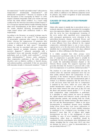
          Figure  1:  (a)  There are five possible areas of ulnar nerve compression   instability. In cases of nerve instability, anterior nerve
          at the elbow level. (1):  Arcade  of Struthers;  (2):  proximal epitrochlear   transposition, either deep or superficial, should be
          region;  (3): epitrochlear‑olecranon channel; and (4): fibrous arch
          between  the humeral and ulnar parts of the flexor carpi ulnaris (FCU),   considered. Notably, nontraumatic nerve debridement and
          better known as Osborne’s arcade; (b) the figure shows the opening of   release,  including that for a long tract  (10‑15  cm), does
          Osborne’s arcade (4) with the release of the septum between the ulnar   not damage  the  nerve  or cause its  devascularization, as
          part of the FCU and the flexor‑pronator group (5). (1), (2) and (3) indicate
          the release of the other areas of decompression     was previously believed. [13,15,23]

          Plast Aesthet Res || Vol 2 || Issue 4 || Jul 15, 2015                                             177

