
Page 62 - Read Online
P. 62
Page 4 of 10 Skladman et al. Plast Aesthet Res 2023;10:66 https://dx.doi.org/10.20517/2347-9264.2022.107
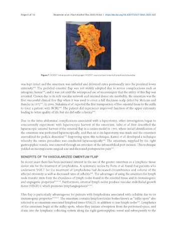
Figure 1. VOLNT Intraoperative photograph. VOLNT: vascularized omental lymph node transfer.
was kept intact and the omentum was unfurled and delivered extra-peritoneally into the proximal lower
[26]
extremity . The pedicled omental flap was not widely adopted due to severe complications such as
iatrogenic hernia , and it was not until the widespread use of microsurgery that the utility of this flap was
[25]
revisited. Chosen due to its rich vascular network and minimal donor site morbidity, the omentum was the
first successful clinical free flap when it was used to cover a full thickness scalp defect by McLean and
Buncke in 1972 . In 2006, Nakajima et al. reported the first transposition of free omental tissue to the axilla
[27]
to treat a patient with BCRL . The patient did experience improved function of the upper extremity
[28]
leading to better quality of life, but she did suffer a hernia .
[28]
Due to the intra-abdominal complications associated with a laparotomy, other investigators began to
concurrently experiment with laparoscopic harvest of the omentum. Saltz et al. first described the
laparoscopic-assisted harvest of the omental flap in a canine model in 1993, where initial identification of
the omentum was performed laparoscopically, and then an 8 cm laparotomy was made and the omentum
exernalized for pedicle dissection . Improving upon this technique, Kamei et al. developed a technique
[29]
whereby the entire procedure was conducted laparoscopically . The omentum, supplied by the right
[22]
gastroepiploic vessels, was removed through an extension of the infraumbilical port incision. This technique
yielded an inconspicuous surgical scar and decreased postoperative pain .
[22]
BENEFITS OF TH VASCULARIZED OMENTUM FLAP
In recent years there has been increased interest in the use of the greater omentum as a lymphatic tissue
donor site for the treatment of lymphedema. A systematic review by Forte et al. found that patients who
underwent VOLT for the treatment of lymphedema had decreased circumference and volume of the
[30]
affected extremity as well as decreased rates of cellulitis . The advantages of using the omentum for lymph
node transfer stem from the abundance of lymph nodes found in the omental tissue and its immunogenic
and angiogenic properties [15,16,31] . Furthermore, omental lymph nodes produce vascular endothelial growth
factor (VEGF) C which promotes lymphangiogenesis [16,31] .
This flap is particularly advantageous for patients with lymphedema associated with cellulitis due to its
immunogenic properties [15,16,31] . The omentum contains lymphoreticular bodies known as “milky spots” also
referred to as omentum-associated lymphoid tissue (OALT), in addition to true lymph nodes . Lymphatics
[16]
of the omentum begin at the milky spots, where they initiate absorption from the peritoneal space, and
drain into the lymphatic collecting system along the right gastroepiploic vessel and subsequently to the