
Page 54 - Read Online
P. 54
Tawaklna et al. Plast Aesthet Res 2023;10:63 https://dx.doi.org/10.20517/2347-9264.2023.40 Page 5 of 9
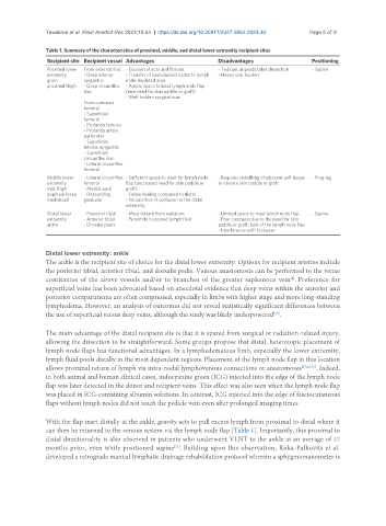
Table 1. Summary of the characteristics of proximal, middle, and distal lower extremity recipient sites
Recipient site Recipient vessel Advantages Disadvantages Positioning
Proximal lower From external iliac - Excision of scar and fibrosis - Tedious, unpredictable dissection - Supine
extremity - Deep inferior - Transfer of vascularized nodes to lymph - Heavy scar burden
groin epigastric node-depleted area
proximal thigh - Deep circumflex - Ample space to inset lymph node flap
iliac (rare need for skin paddle or graft)
- Well-hidden surgical scar
From common
femoral
- Superficial
femoral
- Profunda femoris
- Profunda artery
perforator
- Superficial
inferior epigastric
- Superficial
circumflex iliac
- Lateral circumflex
femoral
Middle lower - Lateral circumflex - Sufficient space to inset for lymph node - Requires debulking of adjacent soft tissue - Frog-leg
extremity femoral flap (decreased need for skin paddle or to obviate skin paddle or graft
mid-thigh - Medial sural graft)
popliteal fossa - Descending - Faster healing compared to distal
medial calf genicular - No sacrifice of perfusion to the distal
extremity
Distal lower - Posterior tibial - Most distant from radiation - Limited space to inset lymph node flap - Supine
extremity - Anterior tibial - Proximity to pooled lymph fluid - Poor cosmesis due to the need for skin
ankle - Dorsalis pedis paddle or graft, bulk of the lymph node flap
- Interference with footwear
Distal lower extremity: ankle
The ankle is the recipient site of choice for the distal lower extremity. Options for recipient arteries include
the posterior tibial, anterior tibial, and dorsalis pedis. Venous anastomosis can be performed to the venae
comitantes of the above vessels and/or to branches of the greater saphenous vein . Preference for
[4]
superficial veins has been advocated based on anecdotal evidence that deep veins within the anterior and
posterior compartments are often compressed, especially in limbs with higher stage and more long-standing
lymphedema. However, an analysis of outcomes did not reveal statistically significant differences between
the use of superficial versus deep veins, although the study was likely underpowered .
[28]
The main advantage of the distal recipient site is that it is spared from surgical or radiation-related injury,
allowing the dissection to be straightforward. Some groups propose that distal, heterotopic placement of
lymph node flaps has functional advantages. In a lymphedematous limb, especially the lower extremity,
lymph fluid pools distally in the most dependent regions. Placement of the lymph node flap in this location
allows proximal return of lymph via intra-nodal lymphovenous connections or anastomoses [27,29,30] . Indeed,
in both animal and human clinical cases, indocyanine green (ICG) injected into the edge of the lymph node
flap was later detected in the donor and recipient veins. This effect was also seen when the lymph node flap
was placed in ICG-containing albumin solutions. In contrast, ICG injected into the edge of fasciocutaneous
flaps without lymph nodes did not reach the pedicle vein even after prolonged imaging times.
With the flap inset distally at the ankle, gravity acts to pull excess lymph from proximal to distal where it
can then be returned to the venous system via the lymph node flap [Table 1]. Importantly, this proximal to
distal directionality is also observed in patients who underwent VLNT to the ankle at an average of 27
months prior, even while positioned supine . Building upon this observation, Roka-Palkovits et al.
[21]
developed a retrograde manual lymphatic drainage rehabilitation protocol wherein a sphygmomanometer is